Chapter 39. Canthal Fixation
Kimberly A. Swartz, BA; Martin I. Newman, MD, FACS; Henry M. Spinelli, MD, FACS
INDICATIONS
There are many manifestations of a malpositioned eyelid, including ectropion, entropion, and retraction. The etiology of these conditions is often the attenuation of canthal tendons, evident in the descent of the lateral canthal tendon from the 10- to 15-degree incline, compared to the medial canthal tendon, seen in young patients, to a coplanar, or inferior position. The two most effective treatments for lax lateral canthal tendons are canthopexy, suitably for the young patient without extensive lid laxity, and canthoplasty, a more powerful treatment suitable for older patients who present with lid laxity, apparent on a snap back or pinch test.
PREOPERATIVE PREPARATION
In consultation, it is important to note any prior oculoplastic surgery. A history of cosmetic surgery can indicate cicatricial eyelid malposition, which may be accompanied by a deficiency in anterior lamella. In this case, a canthopexy or canthoplasty alone will not suffice, and a mid-cheek suspension, free skin graft, or transpositional graft may also be required for full correction.
ANESTHESIA
Monitored anesthesia care (MAC) or general anesthesia can be used for this procedure.
POSITION AND MARKINGS
Preoperatively, the desired position of the canthus is delineated in 3 planes: cephalic-caudal, anterior-posterior, and medial-lateral. Locations are all important, and when only one side is to be addressed, the contralateral side can serve as a guide.
Incision and Exposure
Local anesthetic with epinephrine should be infiltrated using a 27- to 30-gauge needle along the lower lid, and the planned incision. Next, a protective lens may or may not be put in place, depending on the surgeon’s preference. The incision is then made using a no. 15 blade, either along a lateral canthotomy incision or along an upper lid incision, which is often used in the case of concurrent procedures. At this point, division of the inferior crus of the lateral canthal tendon should be carried out to prepare it for lysis.
DETAILS OF THE PROCEDURE
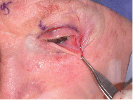
Following severing of the inferior crus of the lateral canthal tendon, the lower lid then must be completely mobilized (Figs. 39-1 and 39-2). This is accomplished by lysing the lateral retinacular structures, including the lower lid retractors and the orbital septum. Once this has been done, a tarsal strip is created by circumferentially deepithelializing a lateral segment of the lower eyelid, which is backcut below the tarsal plate, ultimately isolating approximately 3 mm of distal tarsal plate. This tarsal strip should then be engaged with a double-armed, braided, nonabsorbable suture.

Figure 39-1 At this point, canthotomy and cantholysis are complete, as evidenced by the mobility of the lower lid.
Stay updated, free articles. Join our Telegram channel

Full access? Get Clinical Tree


