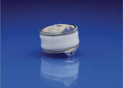
7 The treatment of degenerative cervical disk disorders is one of the most satisfying aspects of a neurosurgeon’s or an orthopedic spine surgeon’s practice. In general, those patients with pathology requiring surgical treatment present with classic findings, complaints, and imaging studies that make the diagnosis of myelopathy and radiculopathy due to disk disease straightforward. The surgical options commonly employed have been extensively studied and their efficacy and safety are well documented. Most importantly, patient outcomes are uniformly satisfying with acceptable morbidity and a mortality rate well below 1%.1,2 In light of the endorsement of the current surgical treatment of degenerative cervical disk disorders, one could reasonably wonder why an entirely new approach such as cervical disk arthroplasty need be considered. The impetus to pursue arthroplasty in the cervical spine is not due to outright failure of current surgical options. Rather, it arises from a desire to avoid the shortcomings and inherent morbidities that accompany those approaches. In the treatment of radiculopathy and myelopathy, ventral removal of compressive disk space lesions is easily achieved with an anterior microdiskectomy. However, subsequent fusion eliminates the motion segment, changing spinal biomechanics in the process. Adjacent segment hypermobility and progressive adjacent segment degeneration have been documented in several reports. Hilibrand et al looked at adjacent segment disease following cervical arthrodesis.3,4 They noted symptomatic disease defined as new onset radiculopathy or myelopathy appropriate for surgical treatment occurring annually in nearly 3% of their patients.3 Radiographic evidence of adjacent segment degeneration can be much higher. Goffin et al found more than 90% of patients treated with fusion had evidence of adjacent segment degenerative disk disease 5 years after surgery.5 Whether or not adjacent segment degenerative disk disease is accelerated by fusion or simply an expression of multilevel degenerative disease has not been conclusively answered in the medical literature. Perhaps tilting the scales in favor of fusion as a significant contributing factor is further consideration of Goffin et al’s paper.5 They compared rates of subsequent adjacent segment disease in patients fused for degenerative disk disease to those undergoing fusion for trauma and found no significant difference. Thus fusion appears to be an independent variable in regard to the breakdown of the adjacent segment because the trauma patients were not undergoing an operation for a degenerative condition. Sagittal plane balance is frequently altered with anterior cervical procedures. Subsidence of the interbody fusion graft and kyphosis of the fused segment are known iatrogenic deformities associated with the fusion procedure.6,7 This loss of lordosis may hasten adjacent segment degeneration because changes to both local and distant intervertebral alignment will alter biomechanics of the spine. Even when fusion does not accompany anterior cervical diskectomy, loss or impairment of mobility of the treated segment is predictable. In one study, ~90% of patients treated with anterior microdiskectomy without arthrodesis went on to spontaneous fusion, with most experiencing kyphotic change.8 Anterolateral foraminotomy has been advocated by a few authors as a method of preserving the disk while providing decompression.9,10 The procedure removes the ipsilateral uncovertebral joint or portions of it to expose and remove the offending pathology. Excellent results have been reported by one surgeon retrospectively reviewing his own results.9 However, the procedure has not enjoyed uniform success. Another report showed that reoperation was required in 30% of the patients treated with that procedure. In addition, the same authors found only 52% of their patients experienced good or excellent outcomes.11 Other morbidity associated with anterior cervical fusion depends on the choices the surgeon makes in regard to technique. Iliac crest graft harvest is associated with chronic hip pain in nearly 30% of patients.12 Plate fixation, currently in vogue, has been shown to contribute to dysphagia, especially when thick or rough-surfaced plates are used.13 Finally, pseudarthrosis is a well-known complication of anterior cervical fusion. Depending on graft material, number of levels fused, and smoking history, a pseudarthrosis rate as high as 50% has been reported.14 The treatment of pseudarthrosis often necessitates a second operation. Another drawback of fusion is the months of activity restriction that most surgeons impose postoperatively on their patients. This has little to do with the recovery from radiculopathy or myelopathy. Rather, this convalescent period is imposed to achieve a solid fusion. Avoiding fusion may be motivation enough to entertain a surgical alternative without even considering the issue of motion preservation. Absent degenerative deformity or instability, fusion is performed primarily to mitigate the iatrogenic damage created during decompression for treatment of radiculopathy and myelopathy. Cervical disk arthroplasty devices are classified according to their makeup. Composed of polymer (polyurethane core) and metal (titanium end plates), the Bryan Cervical Disc prosthesis (Medtronic Sofamor Danek, Memphis, TN) is categorized as a metal-on-poly device (Fig. 7–1). Currently, all other metal-on-poly cervical arthroplasty devices have ultra high molecular weight polyethylene (UHMWPE) cores. UHMWPE has a higher modulus of elasticity than polyurethane, making it less compressible.15 The compressibility of the Bryan core allows it to accommodate sudden axial loads, similar to a shock absorber on an automobile. The dome-shaped, polished titanium end plates are merged with a polyurethane sheath. The sheath prevents fibrous tissue ingrowth and contains particulate debris generated by the moving surfaces. Located inside this shell is the donut-shaped polyurethane nucleus. At the top and bottom of the device are filling ports that allow sterile saline instillation prior to insertion. The saline functions as a lubricant. The device is axially symmetric and also symmetric in a caudad to cephalad direction. Figure 7–1 The Bryan disk device has porous titanium end plates that promote bony ingrowth. Device diameters range from 14 to 18 mm with one height. The metal tabs on the right side of the figure attach to the insertion instrument and also eliminate the risk of device migration into the central canal. Stability of insertion describes the resistance the device has to extrusion before biological incorporation occurs. For the Bryan disk, this is dependent on fixed interspace distraction and a drilling tool that “mills” concavities in the adjacent vertebral body end plates that conform to the device’s dome-shaped titanium end plates. Ligamentous tension from distraction of the interspace combined with the milled vertebral body end plates results in an interference or a “hand in glove” fit providing initial stability. No screw fixation is utilized with the Bryan device. Ultimately, fixation depends on bony ingrowth into the porous end plates. Ingrowth is a feature found solely with the Bryan device. Currently, other cervical arthroplasty devices that rely on biological fixation use a titanium plasma spray that is an ongrowth surface. The Bryan disk is axially symmetric and unconstrained in the normal range of cervical spine motion. It also has a mobile center of rotation like a normal cervical disk. Device flexion and extension of 11 degrees can occur on an infinite number of radii. The device allows for coupled motions of angulation and rotation. These parameters, combined with translation of up to 2 mm, allow the Bryan disk to mimic normal disk function. As mentioned earlier, the Bryan device is also able to absorb sudden axial loads. This is unique among cervical arthroplasty devices. Like all implanted devices, particularly those that incorporate motion, some degree of device deterioration and component debris is expected with cervical disk arthroplasty. With repetitive motion a certain amount of the device’s mass will be shed into the surrounding tissues, with the potential for distant transport as well. Particulate matter is of concern in synovial joint arthroplasty and has been linked to osteolysis and subsequent implant failure. The disk space is classified as an amphiarthrodial joint. It does not have a synovium. Instead, it is connected to bony surfaces by fibrous and cartilaginous tissue only. Because the joint lacks synovium, concerns for a focal reaction to wear debris are not as acute. Although particle toxicity or an immune response to debris is a potential concern, the materials in the Bryan device have a long history of orthopedic and cardiac surgery usage without undue concern about toxicity.16 Debris created from the articulation between the nucleus and the metallic end plates is theoretically contained by the sheath, preventing a biological reaction. Detailed wear analysis within a motion simulator showed that after 10 million cycles the Bryan device lost 0.75% of its mass. Animal studies found debris in the tissues in one of two chimpanzees and four of nine goats. All goats were implanted with the spine in a flexed orientation, creating a worst-case environment for sheath abrasion against posterior bone unique to the goat model. Particle generation and release were achieved, allowing the development team to evaluate particle morphology and biological response. Particles averaging 3.9 m were noted in the loose connective tissue of the epidural space without an inflammatory response. This is in contrast to control group animals with cervical plates that had more debris and inflammatory responses identified.16
Bryan Cervical Disc Device
 The Bryan Arthroplasty Procedure
Bryan Cervical Disc Features
Stay updated, free articles. Join our Telegram channel

Full access? Get Clinical Tree


 Bryan Cervical Disc Features
 Clinical Experience
 Summary


