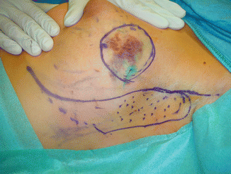
Fig. 10.1
(a, b) Preoperative view. A 36-year-old patient with a 3 cm invasive cancer in the upper outer quadrant of the left breast. The breast was of medium size and moderate ptosis

Fig. 10.2
The tumor was in the upper outer quadrant (circle). The dimensions of the flap to be rotated into the quadrantectomy defect are outlined on the skin
10.2 Surgery
The tumor was excised through an incision in the lateral thoracic wall. This incision was also used for sentinel node biopsy (3 negative sentinel nodes) and flap preparation. Frozen section examination found the tumor completely removed. Due to the wide clear margin toward the skin, no skin overlying the cancer was resected. A flap nourished from perforators from the lateral thoracic wall was dissected off the fascia, de-epithelialized, and rotated into the defect. The donor site defect was closed after adequate mobilization of the tissue.


