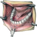
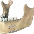
30 Bone Graft and Membrane Stabilization in Implantology Localized repair of severe ridge defects for implant placement using autogenous bone grafts is a very well accepted method (Fig. 30.1). The iliac crest is the most common donor site in maxillofacial reconstruction for the repair of large defects. For the repair of localized severe alveolar defects alternative donor sites such as the mandibular symphysis (Misch, 1992), the oblique line (coronoid) as described by Schliephake (1995, 1996), and the maxillary tuberosity (Moenning and Graham, 1986) are also useful. In small defects, bone augmentation can be performed by guided bone regeneration under membranes. Larger defects require bone grafts, often in combination with membranes or titanium mesh. The fixation of the bone grafts by membranes, titanium mesh, or screws is important for incorporation without resorption. Membranous bone grafts seem to show less resorption than endochondral bone grafts (Smith and Abramson, 1974; Zins and Whitaker, 1983) and revascularize more rapidly (Kusiak, Zins, and Whitaker, 1985). The bone grafts are harvested from the chin region in one or two complete blocks, or from the oblique line (coronoid) in one or two complete blocks, by the use of an oscillating blade or a Lindemann burr and chisels. When the bone is harvested from the maxillary tuberosity, chisels are used. In all techniques it is possible to obtain some cancellous bone chips, which can be used to fill any residual defects. Bone dust and fillings can be retrieved by use of a bone collector in the suction unit, and these can be placed into small gaps.
Introduction
Technique
Related posts:
 Sublingual Hematoma: Pathognomic of Fracture of the Mandible
 Materials and Instrumentation
 Application of Resorbable Plates, Screws and Pins for the Treatment of Midface and Condylar Neck Fractures, and Correction of Craniosynostoses
 Orthognathic Surgery Distance Screws
 Naso-ethmoid Fractures
 Mandibular Fractures Including Atrophied Mandible
Stay updated, free articles. Join our Telegram channel

Full access? Get Clinical Tree


