Benign skin lesions are very common and constitute a large number of skin consultations in general practice. It is important to be able to make a confident correct clinical diagnosis to reassure the patient or to suggest further appropriate management which may include surgical removal.
It is essential that the patient understands the lesion is likely to be benign prior to embarking on a procedure that may lead to a less than ideal cosmetic appearance, hence leading to patient dissatisfaction.
When considering these lesions it is important to think about their derivation (Table 33.1).
Benign Melanocytic Naevi
These consist of three main types: junctional, compound and intradermal melanocytic naevi. These are all benign and are called moles. These are common and appear in childhood and puberty and can become smaller in later life. It is not unusual to see up to 30 naevi in sun exposed areas. However, those with fair skin, a family history of moles and increased sun exposure are likely to have larger numbers (>40).
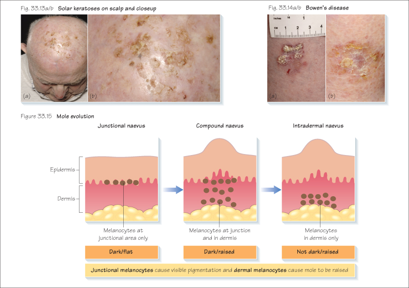
A melanocytic naevus can change through the three stages from a junctional naevus (usually flat and dark) to a compound naevus (raised and usually dark) to an intradermal naevus (usually flesh coloured) (Figures 33.1–33.3 and 33.15).
- As a junctional naevus is flat and brown, a new mole may be difficult to differentiate from malignant melanoma and hence may need to be excised for histology (Figure 33.1). If confident that it is a mole, it can be left alone or excised with a narrow 2 mm margin.
- A compound melanocytic naevus is raised, pigmented and can be hairy. Again, if the diagnosis is confident, it can be left alone (Figure 33.2). A shave excision is better cosmetically than a full ellipse excision for these moles. The patient should be warned regarding the likely cosmetic result of a flat scar and that pigmentation and hairs can regrow within the area.
- An intradermal naevus is usually raised and non-pigmented and needs to be differentiated from a basal cell carcinoma (BCC) (Figure 33.3).
Freckles and Lentigos
Freckles are common benign lesions that usually occur on sun exposed areas and consist of multiple pigmented macules that darken after sun exposure (Figure 33.4). These start in childhood and do not have an increase in melanocytes but the melanosomes within the melanocytes produce increased melanin in response to sun exposure.
A lentigo
Related posts:

Stay updated, free articles. Join our Telegram channel

Full access? Get Clinical Tree


