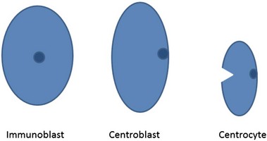
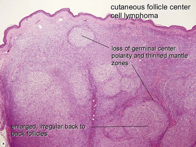
B-cell lymphoma and lymphocytic leukemia

Chapter 25


B-cell lymphoma and lymphocytic leukemia


image


A summary table on ‘WHO Classification of B-cell Tumors of Hematopoietic and Lymphoid Tissues’ and a lymphoma atlas can be found in the on-line content for this book.


Cutaneous B-cell lymphoproliferative disorders


The primary cutaneous B-cell lymphomas share some histologic features with their nodal counterparts, but in many cases represent distinct clinicopathologic entities with significant clinical, immunophenotypic, molecular, and prognostic differences from their extracutaneous counterparts. In general, primary cutaneous B-cell lymphomas have a better prognosis than their nodal-based counterparts, and treatment strategies for them may be different. The classification system used in this chapter reflects the 2005 World Health Organization/European Organisation for Research and Treatment of Cancer combined consensus conference classification for lymphoproliferative diseases of the skin. Note that not all entities included here are necessarily of primary cutaneous origin (e.g. chronic lymphocytic leukemia/small lymphocytic lymphoma, mantle cell lymphoma, Burkitt lymphoma, intravascular large B-cell lymphoma, lymphomatoid granulomatosis) but may frequently involve the skin.


Primary cutaneous marginal zone lymphoma


Primary cutaneous marginal zone B-cell lymphoma is an indolent lymphoma of small lymphocytes of B-cell origin, including centrocyte (marginal zone)-like cells with small to medium-sized slightly irregular nuclei with inconspicuous nucleoli, admixed with plasmacytoid lymphocytes and occasional centroblast-like cells. Marginal zone lymphoma is one of the most common types of cutaneous B-cell lymphoma. The prognosis is excellent, with 5-year survival rates approaching 100%. The lesions usually present as violaceous nodules or plaques preferentially on the trunk and extremities, with upper extremities more commonly involved than lower extremities. The lesions have a nodular or diffuse architecture, often with a characteristic “inverse” pattern relative to benign reactive lymphoid follicles, i.e. they show a dense, darker-staining central nodular portion of benign reactive lymphocytes, which may contain germinal centers, surrounded by neoplastic lymphoid cells with slightly irregular nuclei and more abundant paler cytoplasm (monocytoid or marginal zone B cells). Often, the neoplastic lymphoid population only represents a minority of the cells in the lesion. The epidermis is spared, without epidermotropism of lymphocytes. Surrounding benign reactive germinal centers may be present, and are often colonized by tumor cells. In some cases, the neoplastic cells may surround and infiltrate eccrine coils, similar to the lymphoepithelial lesions seen in extranodal marginal zone lymphomas of the gastrointestinal tract (MALTomas). Primary cutaneous marginal zone B-cell lymphoma includes cases designated primary cutaneous immunocytoma in earlier classification systems. Primary cutaneous immunocytomas are often associated with Borrelia and demonstrate high numbers of monotypic (i.e. they demonstrate monoclonal intracytoplasmic immunoglobulin) plasma cells and lymphoplasmacytoid lymphocytes, some of which may contain intranuclear immunoglobulin deposits (Dutcher bodies).


Primary cutaneous follicle center cell lymphoma


Primary cutaneous follicle center cell lymphoma is also an indolent mature B-cell lymphoma, which exhibits a predilection for the head and trunk. In contrast to its nodal counterpart (primary nodal follicle center cell lymphoma), primary cutaneous follicle center cell lymphoma has a more favorable prognosis, with 5-year survival rates >90%. As with their nodal counterparts, these lesions may have a follicular, follicular and diffuse, or diffuse architecture, most commonly diffuse. However, it must be emphasized again that, despite similar appearances by hematoxylin and eosin, these lesions have different immunohistochemical, genetic, and prognostic features from primary nodal follicle center cell lymphomas. Histology shows a nodular or diffuse proliferation of centrocyte-like lymphocytes with small, slightly irregular nuclei, and variable numbers of larger centroblast-like cells with larger, rounded vesicular nuclei and one or a few prominent nucleoli. In nodular lesions, the follicles appear monomorphous, with a loss of the normal polarity of light and dark zones of the germinal centers, an attenuated mantle zone around germinal centers, and lack of tingible body macrophages in the germinal centers. In larger lesions, the center may show a diffuse architecture, with residual monomorphous neoplastic follicles at the periphery.


Stay updated, free articles. Join our Telegram channel

Apr 26, 2016 | Posted by in Dermatology | Comments Off on B-cell lymphoma and lymphocytic leukemia

Full access? Get Clinical Tree

Get Clinical Tree app for offline access