History of laser use in Otolaryngology – Head and Neck Surgery
Laser safety
Laryngology
Otology
Oropharyngeal surgery
Rhinology
Pediatric otolaryngology
Conclusions
History of Laser Use in Otolaryngology
Historically, in otolaryngology, the laser was used like a scalpel or cautery to incise, cauterize, and coagulate tissue in a precise fashion. Contemporary laser use in the head, neck, and upper airway now focus upon treating for a wide range of clinical applications. By selecting the correct laser wavelength and selecting appropriate dosimetry parameters, surgeons can control both the temporal and spatial evolution of heat within the target site, creating different tissue effects depending upon the desired clinical outcome. Light is variably absorbed and scattered as it propagates through tissue, and the distribution of light in tissue determines the nature of the specific interaction.1 As in dermatology, the selection of the appropriate laser in head and neck applications is critically important. The selection of a specific device and mode of delivery are just as important as wavelength and dosimetry.
The Otolaryngologist – Head and Neck surgeon frequently works in areas that are either difficult to access using classic methods or require extreme precision, and in these circumstances laser technology can be of immense value. For example, during microsurgery of the larynx or ear, a micromanipulator or microscope-mounted scanner, is needed to precise focus and translate a laser beam as small as 100 μm across minute target sites. In surgery of the subglottis and trachea, flexible optical fibers (fiberoptics) or waveguides are used to deliver laser light to these remote locations. In minimally invasive laryngeal cancer operations, high power CO2 lasers are needed to cut tissue while maintaining hemostasis.
Frequently Used Lasers in Otolaryngology
The workhorse in Otolaryngology – Head and Neck Surgery is the carbon dioxide laser. It is used extensively in surgery of the larynx and ear because it can cut tissue precisely, seal small blood vessels and also ablate bone. In cancer surgery, CO2 lasers may seal lymphatics, potentially reducing the spread of disease. Since infrared light is invisible, the CO2 laser is always used with a visible aiming beam. The potassium-titanyl phosphate (Neodymium-doped:Yttrium-Aluminum-Garnet), KTP(Nd:YAG), laser is also used in laryngotracheal surgery as well as surgery of the oropharynx and nose. The KTP(Nd:YAG) laser is most frequently used for vascular lesions because of its absorption by oxyhemoglobin. In the nose, the KTP(Nd:YAG) laser is most frequently used for treatment of hereditary hemorrhage telangiectasias (Osler-Weber-Rendu disease). This is a visible wavelength laser and hence easily transmitted using low-cost optical fibers. It can be made to produce a pulsed output beam, also known as giant pulse laser or Q-switching, that allows for the delivery of nanosecond pulses, which lead to less tissue injury.2 Nd:YAG lasers are used in otolaryngology as well, however the deep penetration depth of this wavelength has resulted in limited use; though in contact mode, this laser has applications as a precise cutting device.3–6
The Argon laser is also used for nasal and middle ear surgery. It is absorbed by hemoglobin and melanin and is generally used in continuous-wave mode. The Holmium:YAG (Ho:YAG) laser is a mid-infrared laser wavelength that is moderately absorbed by water, its principal chromophore. It is valuable because it has a relatively shallow optical penetration depth like the carbon dioxide laser, but can be transmitted using low-cost silica fibers. Its use in otolaryngology has been very limited thus far due to the lack of availability of these devices in most medical centers. The Erbium:YAG laser is used for both nasal surgery, in rhinophyma, and cosmetically for skin resurfacing. It has high absorption in bone and minimal thermally-induced peripheral damage. It also has a tolerable photoacoustic wave effect in the surrounding tissue.7 Pfalz, Fisch and others have developed Erbium:YAG lasers for middle ear surgery, but the high cost of these systems severely limited adoption. There currently is no commercial manufacturer of Erbium:YAG laser systems designed for middle ear surgery (Table 1).
Laser type | Wave length | Penetration depth | Delivery methods | Indications/applications |
|---|---|---|---|---|
Argon | 514 nm | 0.8 mm | Fiber | Ear – stapes surgery |
Micromanipulator | Nose – telangiectasias | |||
Focusing handpiece | ||||
CO2 | 10,600 nm | 30 μm | Articulated arm Micro-manipulator hollow wave-guide scanner Focusing handpiece | Glottis/subglottis/larynx/oropharynx Benign/malignant |
Therapeutic/diagnostic | ||||
Tonsils | ||||
Lingual | ||||
Oral cavity/tongue | ||||
Benign/malignant | ||||
Therapeutic/diagnostic | ||||
Nose | ||||
Turbinate hypertrophy | ||||
Erbium: YAG | 2,940 nm | 3 μm | Articulated arm sapphire fiber | Nose Rhinophyma |
Cosmetic | ||||
Laser resurfacing | ||||
Holmium:YAG | 2,120 nm | 0.4 mm | Fiber/bare fiber contact Handpiece | Nose |
Sinus surgery Turbinate hypertrophy | ||||
KTP (Nd:YAG) | 532 nm | 0.9 mm | Fiber/bare fiber contact Side-fire Focusing handpiece Diffuser tip Micromanipulator | Nose |
Polyps Epistaxis Oropharynx/palatine tonsils Obstructive sleep apnea Vascular malformations | ||||
Nose | ||||
Telangiectasias | ||||
Trachea | ||||
Stenosis | ||||
Subglottic hemangioma | ||||
Nd:YAG | 1,064 nm | 4 mm | Fiber/bare fiber Contact tips | Vascular malformations Tumor removal (contact mode) |
Turbinate surgery |
Laser Safety in Otolaryngologic Applications
As in dermatology, laser safety needs to be considered for the patient, surgeon and surgical staff. The use of a laser near the eyes and in the airway warrants the diligent practice of laser safety at all times. Blindness, burns to the skin, airway injury and death may result if precautions are not taken to prevent these devastating events. The safety precautions and measures to prevent eye injury in head and neck surgery are identical to those used in dermatology, with the exception that the laser dosimetry is often significantly more powerful than that used to treat the skin. Eye protection measures are reviewed elsewhere.
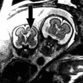
The use of lasers in surgery in the airway presents a major challenge that is unique to otolaryngology – head and neck surgery. Flammable anesthetic gases such as nitric oxide and halothane increase the risk of fire and explosion during airway surgery, particularly when using a high laser power in the presence of flammable endotracheal tubes that may serve as an initiator for combustion in an oxygen rich environment.10 Specialized laser-safe endotracheal tubes (Fig. 1) exhibit prolonged mean times to ignition in comparison to standard endotracheal tubes. Reflective metallic tape wrapped around the endotracheal tube is shown to reduce the risk of ignition of all tubes.11,12
Fig. 1
Laser-specific endotracheal tube wrapped with reflective metallic tape. Laser-Shield II® Endotracheal Tube – Medtronic
Foth has extensively studied the safety of endotracheal tube use in laser surgery. He evaluated the increase in temperature of both the external and internal aspects of a variety of endotracheal tubes that are commonly used in anesthesia. He found that, in comparison to a metallic tube, the temperatures measured in the compound tube were much lower and that a significant difference was observed between the temperatures inside and outside of the compound tube.13–16
Laser Safety Protocol (Adapted from12)
I. Eye protection A. Patient 1. Tape patient’s eyes shut 2. Double layer of saline-saturated eye pads placed over the patient’s eyes B. Operating room personnel 1. Wear appropriate protective glasses with side protectors 2. Surgeon does not require protective glasses when working with the operating microscope or when endonasal using a Hopkins rod endoscope C. Warning signs 1. Placed outside of all entrances in the operative room where the laser is used 2. Caution persons entering the room that the laser is in use and that protective glasses are required D. Limited access 1. Limit traffic into the operating room when the laser is in use 2. Keep doors to the operating room closed when laser is in use |
II. Skin protection A. Use a double layer of saline-saturated surgical towels, surgical sponges or lap pads to cover all exposed skin and mucous membranes of the patient outside of the surgical field 1. Keep this protective layer wet during the case 2. Do not forget to protect the teeth, when exposed B. When microlaryngeal surgery is performed, the patient’s face and peri-oral area is completely draped with saline-saturated towels III. Smoke evacuation A. Aspirate laser-induced smoke from the operative field B. Have two separate suction set-ups available in the operating room 1. One suction for laser-induced smoke and steam 2. One suction for blood and mucous C. Use filters in the suction lines for laser-induced smoke and steam IV. Anesthetic considerations A. Use of nonflammable general anesthetic B. Limit oxygen concentration to maximum of 30% when the laser is in use 1. Mix oxygen with helium, nitrogen or air – do not use nitrous oxide 2. Mix oxygen with helium C. Use a laser-specific endotracheal tube wrapped with reflective metallic tape (e.g., Rusch red rubber tube – Hudson RCI, Temecula, California; Laser-Shield II® Endotracheal Tube – Medtronic Jacksonville, FL) D. Protection of the endotracheal tube cuff 1. Use saline-saturated cottonoids to protect the endotracheal tube cuff (a) Keep the cottonoids moist (b) Count the cottonoids 2. Inflate the cuff with methylene blue colored saline E. Instrument selection 1. Use a wide-bore microlaryngoscope 2. Choose instruments with a non-reflective surface |
Laryngology
Jako and Strong pioneered the use lasers in laryngeal surgery in the 1970s. They were among the first to describe the laser excision of an early laryngeal cancer.17–19 Lasers gained popularity in the 1980s for removal of benign laryngeal lesions such as recurrent respiratory papillomatosis.20 During this time, Steiner began his seminal work expanding the scope of laryngeal laser surgery to treat more extensive malignant tumors of the larynx and upper-aerodigestive tract. His pioneering work is perhaps the greatest contribution to organ sparing laryngeal surgery over the past 20 years and demonstrated the advantages of utilizing microsurgical laser techniques in the treatment of laryngeal cancer and its equivalency to open techniques for specific stage disease.21
The CO2 laser was the first wavelength used in laryngeal surgery and remains the laser of choice for the majority of laryngeal operations requiring this technology. Recent advances in laser technology, anesthesia, and surgical instrumentation have made minimally invasive laryngeal procedures more common. These new approaches have led to better organ preservation rates, improved post-operative functionality and reduced recovery time. It is important to remember that endoscopic examination under general anesthesia is typically performed prior to definitive treatment in order to ensure that comprehensive therapeutic decisions can be made.
Lasers provide surgeons with a precision cutting tool. Using a micromanipulator coupled surgical laser (Fig. 2) at 25× to 40× magnification can negate intention and physiologic tremor of the surgeon and also allow for simultaneous use of both hands and the laser (Fig. 3). The laser can also reach areas that are otherwise difficult to access without performing an open procedure (neck incision, pharyngotomy, laryngectomy, etc.). Presently, commercially available hollow waveguides and fiberoptics continue to expand the role of lasers in airway surgery.
Fig. 2
Micromanipulator coupled surgical laser with patient placed in suspension (Photograph provided courtesy of Dr. Brian Wong)
Fig. 3
Micromanipulator coupled surgical laser. Note the bimanual instrumentation allowed by placing the patient in suspension (Photograph provided courtesy of Dr. Brian Wong)
Indications and Contraindications Indications Malignant Lesions Glottic carcinoma – Early stages Supraglottic carcinoma Hypopharyngeal carcinoma Palliative therapy (tumor debulking) Benign Lesions Polyps Papillomatosis Hemangiomas and Vascular Malformations Laryngomalacia Vocal Cord Paralysis Reinke’s Edema Contraindications Abnormal anatomy that precludes suspension laryngoscopy Malignant Lesions Glottic – Late stages III / IV Extension into surrounding tissues Recurrent disease after radiation therapy Hypopharyngeal carcinoma Benign Lesions Lesions located at the anterior commissure Lesions located at the free edge of the vocal cord Cricoid cartilage stenosis |
Malignancy
Transoral endoscopic laser laryngeal microsurgery depends upon the ability of the surgeon to determine the extent of tumor invasion prior to excision via detailed physical examination performed under general anesthesia and imaging studies. The primary objective is to achieve complete resection of the cancer with clear margins, while maintaining as much organ function as possible (e.g., swallowing, speech, etc.). While transoral laser laryngeal microsurgery (TLM) has many advantages such as decreased post-operative morbidity, reduced recovery time and the potential for organ preservation, recurrence or incomplete resection are significant risks. Hence, the procedure requires skill and sound clinical judgment. It is generally contraindicated in patients with known extensive invasion into other structures and recurrent cancer in previously irradiated areas.22
Glottic Carcinoma
The glottis is the area of the larynx that includes the vocal cords. Use of the laser to excise glottic cancer is well established for lesions staged at T1 and early T2. For all T2a carcinomas of the glottis, laser excision is recommended regardless of the pattern of the spread of the tumor.19,23,24 Provided the tumor is superficial, it is of limited significance whether the disease involves the supraglottis, subglottis or the anterior commissure. The superficial location of the tumor in T1 and early T2 disease allows it to be excised by partial mucosectomy. Laser-assisted endoscopic excision of T3 and T4 laryngeal carcinomas is controversial. Most experts in the field reject the idea of laser microsurgical excision of these advanced tumors unless a classic laryngectomy is not an option due to underlying co-morbid disease. Patients with T3 or T4 laryngeal lesions require the widest possible endoscopic excision followed by post-operative radiotherapy to the primary site and surrounding groups of lymph nodes; however they more often undergo a classic open total or partial laryngectomy.25,26 Steiner groups patients with T3 (n = 17) and T4 (n = 6) lesions in the same category as patients with T2b glottic and supraglottic lesions and routinely treats these tumors with laser-assisted endoscopic surgery.25
Supraglottic Carcinoma
Supraglottic tumors typically occur either in the suprahyoid epiglottis and false cord area or in the infrahyoid/epiglottic area. It is relatively rare that small, well-circumscribed tumors are diagnosed in the supraglottic area because the tumors only produce symptoms, and therefore are brought to the attention of a physician, when they are much larger. Only a few reports have been published on the laser resection of early stage supraglottic carcinomas.27–31 Tumors in the suprahyoid epiglottis and false cord area are easily removed. Laser excision is indicated because wide margins can be taken without the concern of adversely affecting function.
Infrahyoid epiglottic cancers are difficult to assess pre-operatively even with imaging studies and are at risk for infiltrating the pre-epiglottic space. There are conflicting opinions regarding endoscopic laser resection of carcinoma that involves the pre-epiglottic space. Iro urges caution and restraint in treatment of T3 staged supraglottic cancers with transoral laser surgery.32 Whereas Rudert believes that these supraglottic cancers that invade the pre-epiglottic space are candidates for endoscopic laser resection.33 If endoscopic excision is performed and margins remain positive, salvage radiotherapy will not be sufficient, and classic laryngectomy may be required.
Hypopharyngeal Carcinoma
Pre-resection analysis of the airway during surgical endoscopy under general anesthesia may not be able to identify the true extent of hypopharyngeal tumors. Imaging studies can aid in this endeavor. Tumors involving the piriform sinuses usually involve the thyroid cartilage, arytenoids, paraglottic and pre-epiglottic space as well as other soft tissues of the neck with little or no evidence seen on endoscopy. Because of these factors, the majority of laryngologists agree that partial pharyngo-laryngeal resection utilizing only a laser is insufficient for complete eradication of disease and cannot be justified. Classic open surgery (i.e., laryngectomy, laryngopharyngectomy) remains the standard of care.
Palliative Therapy
The laser is a useful tool in debulking tumors that obstruct the upper airway and may be an alternative to tracheostomy.34,35 Avoiding a tracheostomy greatly improves a patient’s quality of life. Palliative airway surgery requires judgment. If too little tumor is resected, the obstruction will persist and ultimately lead to a tracheostomy. However, aggressive resection of tumor may lead to aspiration and the need for a tracheostomy to protect the airway and provide pulmonary toilet. Laccourreye published a 10-year experience describing the use of the CO2 laser to debulk obstructing endolaryngeal carcinomas in 42 patients for the avoidance of a tracheostomy. Ninety-three percent or 39 of 42 patients avoided tracheostomy.36
Recanalizing the upper digestive tract by ablating hypopharyngeal and esophageal outlet tumors poses a serious risk of hemorrhage and will only provide temporary improvement in swallowing. Placement of a percutaneous gastrostomy tube (PEG) tube is a safer and better option for these head and neck cancer patients with dysphagia. If palliative laser surgery is considered, one should aim for sustainable symptomatic relief.
Benign Disease
The CO2 laser has a firmly established role in the treatment of benign laryngeal disorders such as papillomas, polyps, vascular malformations and strictures. Since Jako and Strong first introduced the CO2 laser in 1970, several groups, including those led by Shapshay, Zeitels, Ossoff, Steiner, Motta and Rudert, have published extensively on their experiences with laser surgery in benign laryngeal disease.37 The use of other lasers such as the KTP(Nd:YAG) laser, the pulsed dye laser and the diode laser has also been reported, but their use has not been as widespread due to lack of availability in most medical centers. Therefore, the carbon dioxide laser remains largely the laser of choice.
Polyps
The vocal cords have a delicate subepithelial tissue layer that can be damaged as a result of repetitive collisions and shearing forces. Vocal cord polyps are generally unilateral, sessile and appear to be spherical and well circumscribed. They typically originate at the free edge and on the anterior two thirds of the vocal cord. (Fig. 4) Voice abuse and chronic inflammation from tobacco smoke irritation are common causes of injury to the vocal cord epithelium and lead to damage within the epithelial basement membrane. A large percentage of patients with polyps are heavy smokers or are exposed to a large amount of secondary tobacco smoke. When conservative methods (i.e., voice rest, speech therapy, etc.) fail, treatment is surgical removal. It is important to always send the excised polyps for histology to exclude an early malignancy or to diagnose a benign tumor. For this reason, these polyps should never be vaporized with the laser.
Fig 4
Pulsed 532 nm KTP laser assisted subepithelial resection of fibrovascular mass (hemorrhagic polyp) on the middle 2/3 of the left true vocal cord. The green area is refraction from the laser light (Photograph provided courtesy of Dr. Steven Zeitels)
Papillomas
Laryngeal Papillomas are typically caused by HPV types 6 and 11 and can affect any age group (Fig. 5). The virus causes formation of benign epithelial papillomas of the larynx. Diagnosis is made with flexible fiberoptic laryngoscopy or microlaryngoscopy. Although papillomas most commonly occur on the vocal cords, the larynx, esophagus and trachea must be comprehensively examined during endoscopy because the papillomas can occur anywhere along the aerodigestive system. While the CO2 laser is still used to ablate these lesions, the concern over aerosolization of viral particles has made mechanical laryngeal microdebriders an increasingly more common treatment approach. The recent availability of waveguides for CO2 has lead to renewed interest in the laser for use in office-based laryngeal surgery for papillomas without the need for general anesthesia.38
Fig. 5
Laryngeal papilloma (P) at the level of the glottis (Photograph provided courtesy of Dr. Gupreet S. Ahuja)
Hemangiomas and Vascular Malformations
Subglottic hemangiomas present with persistent cough, hoarseness or stridor, and manifest as reddish, well-circumscribed masses during surgical endoscopy. Surgery involves primarily vaporization and ablation rather than excision.39 The CO2, KTP or Nd:YAG laser is used with low power settings (1–3 W), as high power settings can lead to tracheal perforation, pneumothorax or intractable bleeding. Vascular malformations typically are located in the supraglottic area and are generally asymptomatic. Symptoms of a laryngopharyngeal vascular malformation may include foreign body sensation or mild stridor of unknown origin. Carbon dioxide laser excision or Nd:YAG coagulation is the treatment of choice, though other wavelengths can be used as well with appropriate dosimetry. Use of the Nd:YAG laser has a higher rate of postoperative scarring and stenosis due to the deeper penetration depth of this wavelength, but is extremely valuable in treating large vascular malformations where volumetric heating of large regions of tumor are required. These extensive tumors are more commonly found in the oropharynx and oral cavity.40 Ectatic blood vessels along the vocal cord surface (Fig. 6) may alter the pitch and timbre of the voice and can be problematic for singers and other professionals. They are commonly treated using pulse dye laser and other laser devices.
Fig. 6
Pulsed 532 nm KTP laser assisted photoangiolysis of ectatic blood vessels in a singer (Photograph provided courtesy of Dr. Steven Zeitels)
Laryngomalacia
Stridor in children is most commonly caused by laryngomalacia, and 60–75% of childhood cases of stridor are associated with laryngomalacia.19 Approximately 20% of these cases do not resolve spontaneously and will require surgical intervention. Dysphagia, apnea and respiratory distress are all indicators for surgical intervention. Laryngomalacia will be discussed in a later section along with subglottic hemangiomas.
Vocal Cord Paralysis
Patients that have bilateral vocal cord paralysis may develop severe respiratory distress and airway obstruction from the diminished patency of the glottic airway. Vocal cord paralysis commonly presents following surgery where the recurrent laryngeal nerve is inadvertently injured (i.e., during thyroid tumor surgery). In the immediate post-operative period the patient develops stridor and respiratory insufficiency. Acutely, treatment may involve intubation and tracheostomy. For long-term treatment, CO2 lasers can be used to enlarge the glottic chink and increase airway patency.41 This is challenging surgery because it is important to strike a balance between airway patency and voice quality when enlarging the glottis.
Reinke’s Edema
Reinke’s edema is a reactive sub-epithelial fluid accumulation of unknown etiology; although it is frequently associated with smoking and other activities that produce chronic vocal cord inflammation. Submucosal edema seen on endoscopy is a characteristic feature of the disease. Symptoms of Reinke’s edema include hoarseness and other voice changes. The pulsed dye laser at 585nm wavelength can be used to successfully treat this condition. Certain other disorders can mimic the appearance of Reinke’s edema. For example, Reinke’s edema may mimic the chronic inflammatory changes that are associated with early laryngeal carcinoma. Therefore, the surgeon should have a low threshold to biopsy suspicious lesions.
Contraindications for Surgery of Benign Airway Lesions
There are few contraindications to Transoral Laser Microsurgery (TLM) for benign lesions. One important rule is that surgeons should avoid unnecessary disruption or excision of the anterior commissure and the free edge of the vocal cords. Disruption of the anterior commissure may result in extensive scar formation and an anterior glottic web. TLM is also contraindicated in cricoid cartilage stenosis.42 Most contraindications are related to the relative risks of anesthesia or sedation.
Techniques Pre-Operative Management Imaging and Vocal cord mobility assessment Laser parameters Description of Technique Suspension microlaryngoscopy Laser Delivery / Devices Malignant Lesions Benign Lesions Post-Operative Management |
Pre-operative Management
In the pre-operative evaluation, patients always undergo an indirect mirror or a flexible fiberoptic laryngoscopy examination to determine the extent of the lesion and vocal cord mobility. Video stroboscopy of the vocal cords may be included in the pre-operative evaluation to determine the dynamics of vocal cord movement. Some patients may undergo diagnostic imaging (CT or MRI of the neck) to determine the extent of disease. Vocal cord polyps warrant a trial of speech therapy for vocal coaching and training in proper voice use prior surgery. Patients with carcinoma of the larynx, recurrent papillomatosis and Reinke’s edema must be strongly encouraged to quit smoking.
The most common parameters for the CO2 laser beam using a micromanipulator is a 250 μm spot size with a working distance of 350 mm. Laser power is usually below 8 W. In the superpulse mode, where exposure time is 0.1 s or less, a low power setting of 2–3 W is used. The superpulse mode is useful in achieving maximum ablation through minimal penetration and maximum energy absorption at the surface of interest. There is almost no charring due to the minimal thermal diffusion in the surrounding areas around the target. In preparation for incision of epithelium in cases such as Reinke’s edema, microvascular coagulation is required. The laser is typically set for a 0.05 s pulse of 1 W of power using an unfocused beam.20 Recently, flash scanner technological originally developed for use in dermatologic applications has been re-purposed and adapted for use in laryngeal microsurgery. These devices allow creation of user-defined ablation patterns and cuts under precise computer control. The high cost of these systems has limited the broad adoption of this technology (Acublade-SurgiTouch system, Lumenis, Santa Clara, CA).
Description of Technique
Suspension microlaryngoscopy provides a method in which the surgeon is able to directly visualize the larynx and its surrounding structures. Gustav Killian was among the first to describe suspension laryngoscopy in 1912 and later developed the Killian-Lynch suspension laryngoscope along with Robert Clyde Lynch. Zeitels showed effective use of suspension laryngoscopy in 120 cases in a prospective assessment.43 Suspension allows for bimanual direct laryngoscopic surgery.44 Furthermore, suspension laryngoscopy permits the use of a laryngoscope with a larger bore thereby enhancing exposure. Figure 7 demonstrates the positioning of a patient in suspension, and Fig. 8 shows the normal anatomy of the larynx as visualized under suspension microlaryngoscopy.
Fig. 7
Suspension laryngoscopy (Photograph provided courtesy of Dr. Roger Crumley)
Fig. 8
View of the glottis through the laryngoscope. A arytenoids, E epiglottis, V (black) vocal cords, V (white) vallecula (Photograph provided courtesy of Dr. Gupreet S. Ahuja)
With the patient anesthetized and lying supine, the head is fully extended. A rigid laryngeal endoscope is introduced through the oral cavity and past the epiglottis into the larynx until the desired endolaryngeal structures are visualized. The vocal cords should be clearly visualized from the anterior commissure to the vocal process on both sides. The rigid laryngoscope is then stabilized and secured using a suspension arm. Surgery is performed using a microscope with a 400 mm focal length lens or using a Hopkins rod endoscope inserted thru the laryngoscope bore. There are several instances that make suspension microlaryngoscopy very difficult or even impossible. Abnormal anatomy such as a short, stiff neck, long or protruding maxillary teeth, displaced larynx and lesions or a mass at the base of tongue may limit and prevent suspension laryngoscopy in these patients.42
Suspension laryngoscopy provides a direct airway in which anesthesia may be administered either through an endotracheal tube that is inserted parallel to the laryngoscope or via jet ventilation. This direct airway also provides an unimpeded path for the surgeon to access the vocal cords and other structures in the larynx. The rigid laryngoscope also prevents collapse of the pharynx and hypopharynx that would then obstruct the surgeon’s view. The greatest advantage that suspension laryngoscopy provides is that it allows for a minimally invasive approach to laryngeal surgery where traditional approaches have involved large external incisions and significant morbidity.
Lasers and Laser Devices
Since Jako coupled the carbon dioxide laser to the operating microscope in 1972 for laryngeal surgery, the CO2 laser has been the wavelength of choice. The CO2 laser is used in the majority of operations because of its widespread availability in most medical centers and its efficiency in simultaneously cutting tissue and maintaining hemostasis. Other commonly used lasers in otolaryngology include the KTP(Nd:YAG) laser and the argon laser. In laryngeal surgery, the CO2 laser is generally coupled to a micromanipulator attached to the microscope. This allows the surgeon to use microsurgical instruments and the laser at the same time. It also provides maximum surgical field visualization with high magnification and an unobstructed view of the lesion. The CO2 laser has several advantages over conventional surgical instruments that cut or cauterize in that traditional devices may obstruct the field of view or amplify the surgeon’s intention tremor via a lever arm of up to 20 cm. Furthermore, post-operative edema is less with CO2 laser endoscopic surgery compared to non-laser methods, and thus reduces the risk of post-operative airway obstruction and subsequent tracheostomy. Diminishing the amount of edema also improves the patient’s post-operative swallowing function. The thermal damage zone when using micromanipulators introduces limited artifact into the histological assessment, and it coagulates small blood vessels and seals lymphatic channels thus minimizing the incidence of metastases.20 The diameter of the laser beam, pulse duration and irradiance can be adjusted to produce different tissue effects. Spot sizes between 1 to 4 mm are used for tissue ablation and 0.2–1 mm used for cutting with powers varying anywhere from 2 to 10 W. Some micromanipulators further focus the beam down to an even smaller spot size, and combined with short pulse durations or flash scanner technology, can ablate tissue with minimal to no charring just as in skin resurfacing.45 Morbidity of using the CO2 laser when appropriate is far less than cold surgery, and the cost effectiveness is much greater.46
In certain cases where lesions extend out of the surgeon’s visual field, the CO2 laser can be delivered by a flexible hollow waveguide. However there are limitations with the use of the hollow waveguide due to its limited angulation, the diameter of the laser spot and the significant and variable loss of power during transmission.20 Regardless, recent techniques using hollow fiber technology are evolving.47–51
Malignant Lesions
Glottic Carcinoma
T1 and T2a carcinomas of the glottis are typically resected en bloc and clear margins of 1–3 mm are obtained. However with laser surgery, it is possible to maintain more narrow margins that allow for preservation of vocal function and reduction in post-operative edema.
Transoral Laser Microsurgery (TLM) has yielded good functional results and low rates of recurrence from multiple groups since the 1990 s. Studies have shown that it is possible to preserve the larynx in over 92% of cases with a 5 year local control rate that ranges from 80% to 94%.27,52 Treatment of early glottic cancer with TLM has a distinct advantage over other procedures in that it maintains the availability of all treatment options for patients with local or secondary tumor recurrence including laser re-excision, radiation therapy or open partial laryngectomy.53,54
T2b and T3 carcinoma of the glottis typically involves the vocal cords with supraglottic or subglottic extension. The operative technique using lasers for these tumors involves subdividing the tumor into several pieces for removal. Resection of these tumors may involve the cricoid and thyroid cartilage, the arytenoids, the cricothyroid ligament and any involved laryngeal soft tissue.
The application of laser assisted endoscopic surgery via suspension laryngoscopy draws comparisons to Mohs surgery because resections are driven by frozen section histology. One of the greatest differences between endoscopic laryngeal surgery and cutaneous Mohs surgery lies in the fact that Mohs surgery aims to achieve the best cosmetic result as possible while attempting to remove as little tissue as possible without compromising the tumor resection. In laryngeal surgery, functionality remains essential post-operatively in laryngeal cancer patients. Laser assisted endoscopic laryngeal surgery has potential to allow surgeons to excise cancer piecemeal with frozen section guidance. Frozen section guidance of surgical resections in the larynx is difficult due to the complex geometry of the structures within the larynx, making this radically more complicated than Mohs surgery for cutaneous disease. It is important for the surgeon to carefully label the surfaces of the specimens and work closely with pathologists during surgery.
Ambrosch’s experience of 167 patients undergoing laser excision of T2b and T3 laryngeal tumors showed a 5-year rate of definitive local control to be 87%. The recurrence-free survival rate over 5 years was 62%. None of these patients required a tracheostomy after the primary resection.52 Data is limited to the experience from one institution, but the results indicate that 70% of patients with pT2b and pT3 carcinoma remain free of recurrence following primary resection with the laser. In this study, there was minimal morbidity, and the larynx was fully functional post operatively.52
Supraglottic Carcinoma
T1 and T2 carcinoma of the supraglottis is defined as tumor that has not infiltrated the pre-epiglottic fat, immobilized a vocal cord, or metastasized to a local lymph node. Vaughan first described carbon dioxide laser resection of supraglottic carcinoma. Since then, Steiner, Davis and Zeitels have used TLM for supraglottic resections.21,30,55,56 Laser excision of tumors in this area typically involve extensive dissection and removal of involved muscle, cartilage and even portions of the base of tongue and piriform sinuses in advanced cancer. Tracheostomy is unnecessary in most cases due to limited post-operative edema even after extensive laser dissection. However it should be considered in a procedure with high blood loss or elderly patients with decreased pulmonary function.
There are relatively few reports on laser resection of T1 and T2 cancers in the supraglottis. Ambrosch reports a series of 48 patients with supraglottic T1 and T2 carcinoma in which there was 100% and 89% local control rate for pT1 and pT2 tumors respectively at 5 years. The recurrence free survival rate over 5 years was 83%, and the overall 5-year survival rate was 76%. In patients who experienced a recurrence, none needed a laryngectomy as a secondary treatment.27
T3 carcinoma of the supraglottis treated with laser excision is not a common practice. There are very few reports, but Ambrosch describes a series of 50 patients with pT3 carcinoma treated with laser excision. The definitive 5-year local control rate was 91%; however, 4% of the patients required a total laryngectomy for local recurrence, and 12% of the patients died from cancer related disease.52
Hypopharyngeal Carcinoma
Carcinoma located in the hypopharynx carries the worst prognosis of all upper aerodigestive tract tumors. Reasons for poor prognosis lie in the high rate of local recurrence and the increased likelihood of the presence of metastasis in cervical lymph nodes at diagnosis. Though advances have been made in diagnostic imaging, surgery, radiotherapy, and combined approaches, the mortality rate from these tumors does not reflect any improvement in survival.
Though there are reports of use of TLM in excision of hypopharyngeal cancer, there are few that report any treatment results. The use of lasers in hypopharyngeal cancer treatment is limited but used for function preserving purposes. When operating in the hypopharynx, exposure of the larynx and the piriform sinus is essential. Typical carcinomas of the piriform sinus are resected en bloc in order to assess the extent of local invasion. Safety margins of 5–10 mm are used for piriform sinus tumors (Table 2).
Table 2
Benign lesions
Disorder | Laser treatment |
|---|---|
Polyps | CO2 laser excision |
Papillomas | CO2 laser ablation |
Vascular malformations | Pulse dye laser photocoagulation |
Hemangiomas | CO2 or Nd:YAG laser for coagulation and excision |
Vocal cord paralysis | CO2 laser for enlarging glottic chink |
Laryngomalacia | CO2 laser excision of redundant tissue |
Papillomas
Transoral laser microsurgery of laryngeal papillomas is done with the laser on a low power setting. This allows the surgeon to ablate the disease in a controlled manner that limits disruption of the normal surrounding tissue and ablates only the mucosa affected by the papillomas (Fig. 9). Small islands of healthy mucosa left between the ablated areas promote quicker re-epithelialization.19 In cases where there is bilateral vocal cord or anterior commissure involvement, a conservative approach is advised. It is better to leave small papillomas behind than to have post-operative scarring and synechiae of the anterior commissure, also known as an anterior glottic web, which can lead to voice changes. Corticosteroid administration (3 mg/kg Prednisone) peri-operatively assists decreasing edema after extubation, even if there has been extensive resection. Figure 10a and b show a before and after images of juvenile laryngeal papillomas.
Fig. 9
Pulsed 532 nm KTP laser photoangiolysis and ablation of glottal papillomatosis (Photograph provided courtesy of Dr. Steven Zeitels)
Fig. 10
Papilloma in the pediatric airway, before (a) and after (b) laser excision. P papilloma, V vocal cords (Photograph provided courtesy of Dr. Gupreet S. Ahuja)
Laryngomalacia
Anatomically, the cause of the problem in laryngomalacia is an inward collapse of the supraglottic mucosa during inspiration. If conservative measures fail, the surgical treatment for laryngomalacia is a supraglottoplasty. Bilateral laser incision of the aryepiglottic folds and the arytenoid cartilage region is the treatment if the cause of the stridor is from shortened aryepiglottic folds. If the stridor is caused by inward collapse of the epiglottis during inspiration an epiglottopexy is the surgery of choice. This will be described in more detail in a later section on laser use in the pediatric population.
Vocal Cord Paralysis
The most common laser-based procedure for vocal cord paralysis is unilateral microsurgical laser arytenoidectomy. In Steiner and Werner’s experience, excision of the posterior portion of the vocal cords bilaterally with the microsurgical laser offers several benefits. Most of the vibrating parts of the vocal cords and arytenoids cartilage are preserved preventing aspiration while maintaining good voice quality.19
Post-operative Management
Patients who are otherwise healthy and undergo a limited resection are routinely given a single dose of intravenous steroids, observed in the post-operative care unit and discharged on the same day. Patients who have multiple medical problems or have more extensive surgical excisions, and therefore have a higher possibility for airway compromise, are admitted overnight at minimum for airway observation and scheduled steroid dosing. Post-operative antibiotics are not routinely administered. Strict voice rest for the first 48 h is observed followed by modified voice rest (no screaming, no whispering, limited talking) for the next 2 weeks. Patients are then monitored in clinic with repeat flexible fiberoptic laryngoscopy exams. Any change in voice, difficulty breathing or weight loss is monitored closely and may necessitate repeated exams and procedures.
Adverse Events Injury to teeth, lips, tongue, gingiva on intubation Sensation disturbance of the tongue Edema of the oral mucosa Accidental burns / lacerations to the mucosa Hemorrhage Lingual and hypoglossal nerve palsy Abnormal soft tissue scarring Hoarseness |
In general suspension laryngoscopy is safe for patients as long as the patient has gone through proper pre-operative evaluation by the otolaryngologist and anesthesiologist.42 Complications associated with TLM are more pertinent to the use of the laryngoscope instead of the actual laser itself. Difficult intubation or forceful endoscopic suspension can easily chip, loosen, or fracture teeth if the surgeon is not careful with the orientation of the laryngoscope in the oral cavity. In a study of 339 microlaryngoscopy patients, 75% were found to have small mucosal lesions of the oral cavity, oropharynx and lip.57 Though the injuries were minor, they were a source of major complaints for the patients but resolved over a period of several days. Other complications associated with laser microsurgery include lingual and hypoglossal nerve palsy. Prolonged displacement of the tongue can cause severe contusion and swelling that often leads to dysphagia and sometimes-permanent dysesthesia (sensory disturbance) in the tongue.
There are many general safety considerations involving the use of surgical lasers in surgery that were discussed earlier in this chapter. In particular to laryngeal surgery where high-powered lasers are used in small confined areas with flammable gas, the use of caution cannot be emphasized enough. Stray laser light is not a rare event and is capable of causing burns in unwanted areas. It is recommended practice to cover areas beyond the target site to protect from unwanted burns. Control of oxygen content, inhalation agents, and use of special laser endotracheal tubes in these procedures is important.
Though TLM rarely requires tracheotomy post operatively, it is nevertheless a real possibility in the intermediate and advanced cases. Surgeons must always be aware of certain indicators for tracheotomy. Prolonged compression of the tongue during TLM can cause lingual edema that can lead to airway compromise. Sudden secondary hemorrhage is a major risk in TLM and often necessitates a tracheotomy. Large arteries and vessels should not only be cauterized but clipped as well to avoid sudden secondary hemorrhage.
TLM typically causes the formation of large granulomas if cartilage is exposed by the surgical procedure. These granulomas are perpetuated by small osseocartilaginous sequestrae unless they are removed.22 TLM involving the anterior commissure often results in a round anterior glottic effect, and this often leads to breathiness and hoarseness.58
Laser surgery for benign lesions in the larynx is relatively safe compared to most other treatment modalities. It is a minimally invasive technique with almost no complications attributed to the laser itself.
Future Directions Omni Guide Beampath Tissue welding Office based procedures with lasers Femto-second lasers |
Future directions of laser use in laryngology involves new devices for delivery, office based laser applications, the use of the laser for welding tissue and the creation of lasers that are even more precise than those which are currently available. A recent technological advance (Omni Guide, Boston, MA) has provided surgeons the use of CO2 lasers through a flexible fiber. The Omni Guide Beam Path uses a hollow “photonic band gap fiber.” The fiber is placed in a handpiece coupled with a suction that eliminates the need for a micromanipulator and allows the surgeon to have the freedom of a laser cutting tool in his or her hand. The Omni Guide Beam Path has proven useful in the surgery of both benign and malignant lesions.59
Awake office based laser surgery is gaining popularity in the treatment of benign laryngeal lesions because of the relative safety of lasers and the quick recovery period post procedure. Though the CO2 laser has traditionally been the laser of choice for laryngeal surgery, different lasers have been reported to be useful in office-based surgery. In the office, new technologies such as distal chip endoscopy and rare-earth doped fiber lasers have allowed for creative and innovative surgical techniques. Typical diseases treated with these new techniques are dysplasias and papillomas. A 585 nm pulsed dye laser was originally the laser of choice for vascular lesions; however the 532 nm pulsed KTP laser has proven to be superior. The use of the 2,013 nm Thulium laser which mimics the CO2 laser closely may become useful as an office based laser as well.60–64 Even though otolaryngologists are performing more office procedures that incorporate lasers, the breadth of procedures remains limited for important reasons such as airway compromise, which is poorly handled in the office setting.
Otology
Indications and Contraindications Indications External auditory canal lesions Eustachian tube dysfunction Perforated tympanic membrane Exostoses Debulking of Tumors Acoustic Neuroma Cholesteatoma Resection Otosclerosis Vertigo Facial Nerve Paresis / Paralysis Contraindications Anatomic, clinical and physiologic limiting factors Large skull base tumors |
Laser applications in otology include the treatment of vascular lesions in the external auditory canal (EAC), exostoses (bony outgrowths) in the EAC, the debulking of inoperable tumors, Eustachian tube dysfunction, myringotomy/tympanostomy in otitis media, graft fixation in tympanoplasty, stapedectomy/stapedotomy in otosclerosis, tympanosclerosis, removal of cholesteatoma, cochleostomy, labyrinthectomy in benign paroxysmal positional vertigo, endolymphatic hydrops and facial nerve decompression.65
The use of the laser in middle ear surgery is one of the most elegant examples of laser technology use and applications. Perkins pioneered the use of the argon laser in middle ear surgery in the late-1970s performing the first laser stapedotomy for otosclerosis. One of the first uses of the laser in the field was in patients with otosclerosis. The disease is now one of the most common indications for laser use in otologic surgery. Otosclerosis is a localized disorder of bone resorption and deposition (remodeling) in the middle and inner ear that causes progressive unilateral or bilateral conductive hearing loss that typically begins in the third to fifth decade of life. This defect in bone remodeling in the vicinity the stapes footplate and oval window leads to reduced mobility of the stapes and hearing loss. Mechanical causes of hearing loss, also known as conductive hearing loss, can be entirely corrected through surgery.66,67 Patients with otosclerosis benefit from the argon laser through its precise vaporization of the stapedial tendon, mobilizing the posterior crus of the stapes and in stapes footplate fenestration.68,69 This procedure is called a stapedectomy or stapedotomy, and is performed in order to recreate ossicular chain mobility and decrease conductive hearing loss. Knowledge of middle ear anatomy is critical in obtaining good post-operative results with minimal complications. Avoidance of injury to the facial nerve and the chorda tympani nerve is the standard of care.65,68,70–83
DiBartolomeo described other uses of the argon laser in the field of otology. He utilized this laser in the tympanoplasty (repair of the tympanic membrane), stapedectomy, lysis of adhesions and myringotomy (placement of a hole in the tympanic membrane). He describes the “spot welding” technique of the laser that allows for the adherence of a fascial graft placed in substitution of the tympanic membrane onto the soft tissue annulus.84 The use of the argon laser in otologic surgery is precise, safe and effective. Delivery of a laser to the ear and temporal bone can be either via a fiber (KTP and argon) or via a micro-manipulator such as those used with CO2 lasers.65
Eustachian tube dysfunction is a common ear disorder in which patients may experience chronic recurrent ear infections or difficulty in clearing a blocked sensation of their middle ear. A laser myringotomy may be performed to equalize the pressures between the middle and outer ear, and to assist the ear in draining fluid.85–89 Laser Eustachian tuboplasty (LETP) is the practice of utilizing a diode or an argon laser to vaporize select areas of hypertrophic mucosa and submucosa tissue along the length of the Eustachian tube. In a 2-year follow-up study of 13 adults, Poe et al. found that medical management combined with LETP on a select group of patients can be successful in eliminating chronic middle ear effusions.90
A cholesteatoma is a collection of keratinized epithelium that is located in an abnormal location at the external auditory canal, middle ear, petrous bone or mastoid. It is either congenital in etiology or is acquired due to repeated infection. It is not invasive, but it is destroys the bones of the middle ear leading to a conductive hearing loss. It can also erode the mastoid bone and tegmen can possible lead to a cerebral spinal fluid leak. Therefore, the cholesteatoma needs to be removed in its entirety.91 Excision is associated with a high rate of recurrence. The use of a laser in this surgery may assist the surgeon in removing the mass in more difficult to reach areas of the middle ear and mastoid.92,93
Contraindications to laser use for otologic applications include performing a stapedectomy in the presence of active otitis media, an only-hearing ear that responds well to amplification, vertigo and endolymphatic hydrops, certain inner ear malformations or an overhanging facial nerve that completely obstructs access to the oval window niche. Laser stapedectomy in patients with a perforated tympanic membrane must be postponed until the perforation is fixed. In the case of an overlying facial nerve, preservation of the facial nerve is most important, and conventional instruments are used to perform the operation rather than the laser.
Techniques Pre-Operative Management Laser Selection Laser Doppler Vibrometry Description of Technique Stapedectomy / Stapedotomy Cholesteatoma Excision Myringotomy / Tympanostomy Tympanoplasty Labrinthectomy Post-Operative Management |
Pre-operative Management and Laser Selection
Pre-operative management of patients undergoing laser assisted otologic surgery includes a full history and physical, an audiogram and likely non-contrast computer tomography (CT) imaging of the temporal bone and internal auditory canal. When choosing a laser to use near a neurosensory organ like the ear, it is important to consider the potential collateral damage. The thermal and photoacoustic effects in laser use are a function of dosimetry and tissue optical and thermal properties. Bone has substantially smaller water content than skin or other soft tissues and visible wavelengths are not well absorbed. In the absence of any distinct chromophore, using an argon or a KTP(Nd:YAG) laser to ablate bone requires the use of an initiator. An initiator absorbs laser light and undergoes pyrolysis. In middle ear surgery, a small droplet of blood or charred tissue placed on the laser target serves this purpose. Accordingly, there is a risk of thermal injury, as considerable temperature elevations occur within this region, and ablation occurs with at best modest thermal confinement.82 In contrast, infrared wavelengths are well absorbed by both the hydroxyapatite crystals in bone and the interwoven collagen fibers. Ablation proceeds by the classical mechanisms just as in skin, though photoacoustic transients may be generated leading to audible pops. These mechanical transients may propagate through the inner ear and may result in injury to the delicate neuroepithelium.
In general, shorter laser pulse durations lead to less thermal injury provided the conditions for thermal confinement are met.94,95 However, repeated pulses in rapid succession may lead to the build up of heat in the target site; hence successive pulses should be separated by a sufficient amount of time to allow for complete thermal relaxation. Short pulse laser systems (e.g., Erbium:YAG) generate an acoustic pressure wave that leads to a vibration of the ossicular chain which may result in “noise trauma”. The heat generated by the laser may also cause “convection currents” that also lead to vibratory stimulations. Both of these stimulations can cause acoustic injury and should be controlled by appropriate dosimetry selection. Regardless, the mechanical transients produced during laser ablation are comparable or even less harmful than the vibratory effects produced using conventional methods to perform surgery.7,65,82,96,97
Description of Technique
In addition to the argon laser, the CO2 laser, the KTP laser and more recently, the erbium-YAG laser are used to perform a stapedectomy or stapedotomy in patients with otosclerosis. In the stapedectomy and stapedotomy, the stapedial tendon is vaporized with the laser, then the incudostapedial joint is disarticulated using mechanical instrumentation, and the posterior crus is severed with the laser (Fig. 11a, b). The anterior crus of the stapes is then fractured manually without the laser and the stapes superstructure is removed. The laser is then used to place a single large hole or a set of smaller holes generating a “rosette” type pattern in the stapes footplate to accommodate the prosthesis (Fig. 11c). A mobile prosthesis is put into place.65,68,70–83 No study to date has demonstrated which laser among the three most commonly used in stapes surgery (argon, KTP and CO2) is best. Recently, erbium:YAG lasers have been developed for use in performing this operation, however the high cost of these lasers has limited broad adoption.71,98 The success of laser stapes surgery is based on audiometric analysis (hearing tests) and complication rates. Audiometric results after laser stapedotomy in relation to mean differences in the bone conduction auditory thresholds show improvements of 0.53–5.6 dB in those thresholds regardless of the laser system utilized.70,99
Fig. 11
(a) In stapedectomy, the stapes tendon (blue dashed line) is vaporized with the laser and the incudostapedial joint (black dashed line) is disarticulated. The posterior crus (yellow dashed line) is then severed with the laser. (Photograph provided courtesy of Dr. Hamid Djalilian). (b) Intraoperative image of an argon laser directed at the posterior crus of the stapes after having displaced the chorda tympani nerve from the line of fire. (Photograph provided courtesy of Dr. Hamid Djalilian) (c) Intraoperative image of the oval window after laser stapedectomy. Notice the charred appearance of where the stapes was located (yellow dashed line). Also note the preservation of the chorda tympani nerve (black dashed line). The long process of the incus (blue dashed line) is still intact above and will be used to anchor the stapes prosthesis (Photograph provided courtesy of Dr. Hamid Djalilian)
Smaller cholesteatomas and residual tissue in previously attempted excisions of cholesteatoma have been vaporized with the CO2, Argon and KTP lasers with some reports of lower recurrence rates.65 Laser use is particularly beneficial when removing cholesteatoma from the delicate bones of the middle ear such as the stapes or in removing disease that traverses the obturator foramen of the stapes. When using the laser in this application, its energy is absorbed more by the cholesteatoma than by the bony surroundings.92,93
Related posts:


Stay updated, free articles. Join our Telegram channel

Full access? Get Clinical Tree


