Vascular lesions
Port-wine stains
Superficial hemangiomas
Telangiectasias
Spider angiomas
Pyogenic granulomas
Facial angiofibromas
Lymphangiomas
Pigmented lesions
Café Au Lait
Small congenital nevi
Nevus of ota
Nevus of ito
Lentigines
Becker’s nevus
Other conditions
Viral warts
Molluscum contagiosum
Hypertrophic scars
Morphea
Hypertrichosis
Vitiligo
Acne and acne scarring
Lasers for Vascular Lesions
The flashlamp- pulsed dye laser (PDL) has revolutionized the treatment of vascular lesions in children. The PDL’s well-established safety profile makes it the treatment of choice for vascular lesions in the pediatric population. It uses 577–600 nm wavelengths to target the last oxyhemoglobin peak within the blood vessels. PDL has been used successfully to treat vascular lesions such as port-wine stains, superficial hemangiomas, telangiectasias, angiofibromas and pyogenic granulomas; and non-vascular conditions in children such as viral warts, molluscum contagiosum, hypertrophic scars and others. Adverse effects although rare include dyspigmentation, scarring, infection and pain. |
Oxyhemoglobin, the target chromophore in blood vessels, has absorption peaks at 418, 542 and 577 nm and thus the best suited lasers for treatment of vascular lesions emit light in this range (See Table 2). The earliest lasers used to treat vascular lesions in children were the quasi-continuous lasers such as the Argon pumped tunable dye, copper bromide and copper vapor lasers but these have since fallen out of favor due to the high risk of hypertrophic scarring and textural changes.1 In 1989, the flashlamp-pumped PDL revolutionized the treatment of vascular lesions, and today it is the treatment of choice for some hemangiomas, port-wine stains, and telangiectasias in children.2 It uses a flashlamp to energize rhodamine dye and generates a pulse of yellow light. Today’s most commonly used PDL systems emit wavelengths of 585 or 595 nm and are used not only for vascular lesions but also to target blood vessels in non-vascular lesions in children. More recently developed PDLs have longer wavelengths (595 and 600 nm), larger spot sizes (10–12 mm), and higher peak fluence potential, allowing for better treatment of deeper vessels in hemangiomas and port-wine stains. They also have longer pulse durations (1.5–40 ms), which allows for the targeting of larger vessels in more complicated lesions. As long as the pulse duration is shorter than or equal to the thermal relaxation time of target vessels, the laser can achieve selective destruction of target vessels without damage to the surrounding normal tissue. Larger blood vessels have longer thermal relaxation times (TRT) and thus longer pulse durations can be used. In addition, certain PDL systems have excellent dynamic cooling devices which reduce epidermal damage and intraoperative pain.3
Table 2
Vascular lesions
Lasers used to treat vascular lesions in children | |
|---|---|
Quasi continuous lasers | |
Argon pumped tunable dye | 577–585 nm/yellow |
Copper bromide | 578 nm/yellow |
Copper vapor | 578 nm/yellow |
Krypton | 568 nm/yellow |
Pulsed lasers | |
Flashlamp-pumped pulsed dye laser (PDL) | 585 nm/yellow |
Long pulsed dye laser (LPDL) | 595 nm/yellow |
Potassium titanyl phosphate (KTP) | 532 nm/green |
Other lasers such as the 532 nm KTP, 755 nm alexandrite, 1,064 Nd:Yag or Intense Pulsed light (IPL) are used less frequently, for PDL resistant lesions and have less favorable outcomes.
Indications and Contraindications
Indications Port-wine stains Superficial hemangiomas of infancy in the proliferative or involution phase, those encroaching upon vital structures or those that are cosmetically disfiguring Ulcerated hemangiomas of infancy Telangiectasias Pyogenic granulomas Facial angiofibromas Verrucae vulgaris Molluscum contagiosum Contraindications Patients with unrealistic expectations Patients who form scars will be at a greater risk for this post-operative complication Deep or mixed hemangiomas of infancy Pyogenic granulomas wider or higher than 5 mm |
Port Wine Stains
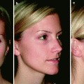
Port-wine stains (PWS) are benign vascular malformations composed of ectatic capillary-sized blood vessels in the papillary dermis that are usually present at birth.4 They occur in 0.3–0.5% of newborns. Early in life, PWS are pink and macular (See Fig. 1)
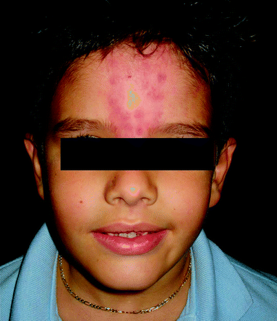
. After puberty some become darker, resembling the color of “port wine,” and hypertrophy. They are usually unilateral and are commonly found on the face. The flashlamp-pumped PDL is the established treatment of choice for PWS in children. One older study used Candela® flashlamp-pumped PDL with a 585 nm wavelength in 89 patients ages 0–31 years of age and compared lightening among the different ages. Using a colorimeter, they measured lightening after five treatments in four different age groups. Good lightening was seen in all age groups and the authors concluded that PDL treatment can be effective at any age.5 However, data from more recent studies using the Candela® V-Beam 595 nm PDL have shown that early treatment of PWS can leads to improvement of the lesion with minimal side effects. The earlier the treatment, the lesion size is proportionately smaller and the thickness of skin is lesser, allowing deeper penetration of the light.6 Starting the treatment early may also require fewer treatments7–9 and may help to increase the chances of complete resolution of the lesion.10 One study reported that treating PWS in children younger than 1 year old seem to have the most effective lightening.11 In addition, aging PWS become thicken and develop nodularity, which can lead to asymmetrical deformity of the face. Once hypertrophy and nodularity occur, the lesion then become very difficult to treat and might not response to PDL.
Fig. 1
Post-treatment purpura secondary to blood vessel destruction typically resolves within several days
Many clinicians have recommended that the decision of when to initiate PWS treatment should be based on patient’s psychosocial benefit, discomfort and anxiety from the procedure. When they are on the face or other cosmetically significant location, PWS can have a profound effect on a child’s social and psychosocial adjustment. For this reason, many physicians have suggested that treatment of PWS is a medical necessity.12,13 According to a psychology study, children develop their body self-awareness around the latter half of the second year of age.14 The development of body self-awareness in toddlers is the foundation of ego development and self-esteem later in life.15 Many families choose to begin treatment before children start school to reduce their psychosocial impact. Studies have shown that adult patients with PWS have lower self confidence,16 which is increase after a significant improvement of PWS has achieved with PDL treatment.17
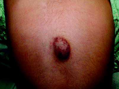
So far, there is no recommended setting of PDL due to the lack of controlled studies with a single parameter difference. In general, the ranges of parameters are: 585–600 nm wavelength, 4–12 J/cm2 fluence, 0.45–10 ms pulse duration and a minimum of 7 mm spot size.1 Longer wavelengths and larger spot sizes may allow the laser beam to penetrate deeper into the lesion.18 Pulse durations should be chosen according to the presumed size of the blood vessels within the lesion. Do keep in mind that capillaries have a TRT in the tens of microseconds and PWS venules have a TRT of tens of milliseconds.1 Post-treatment purpura secondary to blood vessel destruction, is a desired result, and typically resolve within several days (See Fig. 2).19 Many clinicians choose the initial fluence based on their personal experience and adjust the fluence based on the PWS appearance immediately after the laser pulse.20 Some physicians prefer to perform a test spot and gauge PDL settings based on the patient’s response. Using a PDL system that has a cooling device can help reduce epidermal damage and intraoperative pain. PDL with a cryogen cooling spray device was found to enhance clinical efficacy because higher fluences could be used without an increased risk of permanent scarring or dyspigmentation.21
When treating PWS, it is recommended to target the smaller blood vessels and the edges of the lesion first and leave the larger blood vessels for later treatments.4 Multiple treatment sessions are necessity and should be scheduled at 4–8 week intervals. There is no agreement on how many treatment sessions required to achieve complete clearance of the lesion. For facial PWS, the average number of sessions is between 9 and 12 in order to achieve ∼80% clearance.13 The most drastic improvement is noted after the first five treatments. Predictive factors for improvement have been examined by several studies. A study of 91 children with an average age of 4.5 years found that the maximal improvement is noted in the first five treatments in all patients regardless of age, location or size of the lesion. The best result of treatment was observe when treating lesions over bony prominences of the face such as the central forehead, size less than 20 cm2 and those in children less than 1 year of age.11 Lesions of the head and neck require fewer treatments than those on the trunk and extremities and those in the V2 dermatomal distribution of the trigeminal nerve lightened significantly less with the same number of treatments than those in the V1, V3 and C2/C3 regions.22 One study in 16 children recommended that using higher fluences (11–12 J/cm2) and longer pulse durations (1.5 ms) can achieve faster lightening; >75% clearance was attained in four treatment sessions.23 The number of treatments is often determined by when the patient and/or family are cosmetically pleased with the result, or decide not to continue the treatment.
Challenging PWS with larger and deeper blood vessels tend to require more treatment sessions. Repetitive treatments, even when greater than 20 does not increase the risk of side effects and can offer additional lightening.24 Very resistant PWS not responded to repetitive treatments with PDL may require longer wavelength lasers such as 595–600 nm LPDL, 755 nm Alexandrite, 1064 Nd:Yag, KTP and IPL. A recent study demonstrated that KTP laser can help to further lighten PDL treatment-resistant PWS. Of 30 adult and pediatric patients in the study all who have had at least five PDL treatments, 16 patients (53%) experienced some lightening (>25%).25
Hemangiomas of Infancy
While PDL treatment for PWS is well established, the role of PDL in the treatment of hemangiomas of infancy (HOI) is less clear. Hemangiomas are the most common benign tumor of infancy and are composed of capillary-sized blood vessels.1 HOI are estimated to affect 1.1–2.6% of newborns. Their natural history is characterized by three stages: rapid proliferation (usually the first 6–8 months of life), plateau and involution. The duration of each stage in a particular lesion is difficult to predict, however the onset of involution is indicated by a color change from bright red to gray or purple. The vast majority of hemangiomas undergo spontaneous involution without residual defect. On the other hand, about 5–13% of hemagiomas will ulcerate in the proliferative phase and some leave a yellowish discoloration, atrophic scarring, redundant skin, fibrofatty tissue or residual telangiectasias. Treatment of HOI includes active nonintervention, topical, intralesional and oral corticosteroids, vincristine, recombinant interferon alpha 2a and 2b, imiquimod, cryotherapy, and surgery. More recently, vascular lasers such as the PDL have been added to the armamentarium. Due to their unpredictive growth of the lesion and spontaneous resolution of hemangiomas, the indication to treat is controversial. Most experts agree that hemangiomas in locations that is functionally disabling or life threatening (See Fig. 3
), that will permanently scar, located on the face and ulcerated should required treatment. Most pediatric dermatologists prefer a wait and see approach with uncomplicated HOI that do not have the aforementioned characteristics.
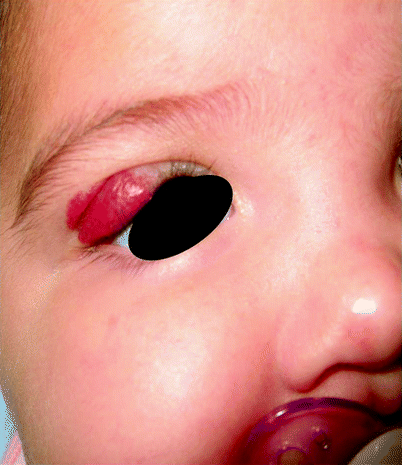
Fig. 3
Deeper hemangiomas may be less amenable to treatment with lasers
Fig. 2
Hemangioma of infancy involving upper eyelid may threaten function and therefore require treatment
Batta et al. compared PDL treatment of uncomplicated HOI to active nonintervention and concluded that there was no useful benefit of early treatment with PDL in uncomplicated hemangiomas. Furthermore, lesions that were treated with PDL experienced an increased risk of skin atrophy and hypopigmentation compared to untreated hemangiomas.28 This study, however, was criticized for not using a cooling device29 which not only allow the use of higher fluences but also prevented post treatment pigment alteration.
PDL treatment is currently used for thin, superficial lesions, ulcerated hemangiomas and the residual redness and telangiectasias left after HOI involute.30,31 LPDL versus traditional PDL may help to limit the average time period of maximum hemangioma proliferation with less adverse effects.32
Clear beneficial results have been seen when PDL is used to treat an early pink macular lesion. One study showed that treating early hemangiomas with 595 nm PDL at fluences of 6–7 J/cm2 can induce involution and prevent the development of the deep components.33 Unfortunately, most HOI do not present to a dermatologist until they are in the proliferative phase.
Clear benefits of PDL are also seen with superficial hemangiomas. In one study, lesions less than 3 mm thick achieved resolved quicker than thicker lesions (>3 mm). However, these are also the ones more likely to involute spontaneously with a good cosmetic outcome. The authors recommended that only superficial hemangiomas impinging on vital structures should be treated with the PDL.33 Furthermore, superficial hemangiomas in proliferative and involution phases both respond well to PDL therapy.34
In addition, PDL treatment in ulcerating hemangiomas can effectively decrease pain and improve healing of the affected area.35 In a study of 78 patients with ulcerated hemangiomas, 91% of patients experienced epithelialization of the ulceration after an average of two laser treatments. No significant complications were noted in this study.36
When treating hemangiomas, it is important to recognize how HOI differ from PWS. As opposed to PWS, hemangiomas are high flow tumors, and composed of smaller blood vessels that close to skin surface.37 Lower fluences and shorter pulse durations should be used in PDL treatment. The average parameter used in PDL treatment for hemangioma studies are: 585–595 nm wavelength, 5–10 J/cm2 fluence, 0.3–0.5 ms pulse duration and a 3–7 mm spot size. Treatment sessions are usually repeated every 1–4 weeks. Kono et al. studied superficial hemangiomas in preproliferative or early proliferative stages in patients 1–3 months of age and compared treatment with a 585 nm PDL (7 mm spot size, 6–7 J/cm2, 0.45 ms pulse duration, no epidermal cooling device) with a 595 nm LPDL (7 mm spot size, 9–15 J/cm2, 10–20 ms pulse duration with an epidermal cooling device). Both groups were treated at 4 week intervals until the lesion cleared. Although the number of children whose lesions showed complete clearance or minimal residual signs at 1 year of age was similar in both groups, the PDL-treated group had more dyspigmentation and more textural changes than the LPDL-treated group. The average time of maximal proliferation in the LPDL group was significantly shorter than in the PDL group. The authors concluded that LPDL is safer and more effective than PDL in the treatment of early hemangiomas.32 However, it is difficult to know which factor confers superiority of the result in LPDL group; the longer pulse duration, the higher fluence, or the use of the cryogen cooling spray.
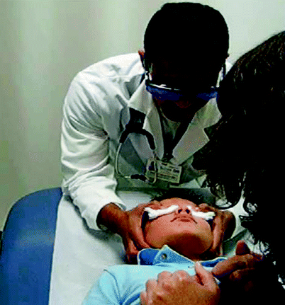
While the efficacy of PDL is limited to treat deeper hemangiomas (See Fig. 4), 1,064 nm Nd:YAG laser has been used successfully to treat PDL-failed lesions.
38 Several studies have used both percutaneous and intralesional applications of the laser with positive results. Nd:YAG laser should only be used by experienced laser surgeons. More recently a dual –wavelength laser system (595 and 1,064 nm) (Cynergy with Multiplex®, Cyanosure, Westford, MA) has been found to be superior than PDL or Nd:YAG alone in clearing facial telangiectasias.39 Combining the 595 nm PDL and the 1,064 nm Nd:YAG wavelengths results in a synergistic effect and would theoretically be beneficial for deeper vascular malformations in children. Studies with this laser in children are ongoing.
Fig. 4
Overlapping 2 × 2-in. gauzes may be held in place over the child’s eyes and may be an alternative to standard safety goggles
KTP laser is a modified Nd:YAG that emits 532 nm through a KTP crystal. Pulse durations range from 1 to 100 ms. The laser is administered to tissue via a fiberoptic hand piece. Advantages of using 532 nm wavelength in treatment of hemangiomas include selective absorption by hemoglobin with minimal purpura. Disadvantage includes the limited penetration depth and the higher risk of post treatment dyschromias.1 Achauer et al. demonstrated successful use of the KTP bare fiber at 15 J and 0.6 mm spot size in treating bulkier hemangiomas.40
Although successful treatment of superficial hemangiomas is evident, there are some controversies about an optimal laser treatment and treatment indication. Clinical treatment criteria include rapid enlargement of the lesion, involvement of large surface area, actual or potential functional impairment, ulceration, cosmetic disfigurement, and recurrent bleeding or trauma to the area.41 Laser treatment should be considered when the lesion failed conventional medical therapy.42 Superficial hemangiomas respond quite easily and effectively to the PDL, while a more variable response is noted in deeper hemangiomas, early proliferative lesions, and ulcerated hemangiomas.33,43
Other Lesions Amenable to Vascular Laser Therapy
In addition to PWS and hemangiomas, other vascular lesions in children can be effectively treated with vascular laser. Telangiectasias are small dilated capillaries, most commonly seen as spider telangiectasias in children.4,44 Telangiectasias, especially on the face, are highly responsive to laser therapy. Complete resolution of the lesion can be achieve with a single treatment in some cases.45 Pyogenic granulomas are solitary vascular proliferations that can be seen in childhood, typically present as rapidly growing, bright red papules. PDL has been found to be more effective for smaller, flatter lesions and should be used to treat the lesion smaller than 5 mm.46 When pyogenic granuloma is not responding to PDL treatment, alternative diagnoses such as spitz nevi should be considered.
There are a growing number of other conditions in children that can be successfully treated with PDL. For example, angiofibromas may be treated effectively when they are first developed and small. The lesions may require follow-up treatments at 6- or 12-month intervals.47 Several studies on the use of PDL in verrucae vulgaris (VV)48 and in molluscum contagiosum49 have shown it to be a useful treatment. VV, or common wart, is estimated to occur in 10–22% of children and young adults with a peak incidence between 12 and 16 years of age.50 Nearly two-thirds of warts resolve spontaneously within 2 years. There are many treatment options for VV including topical salicylic acid, cryotherapy and PDL, which recently been added as a third line treatment of choice for recalcitrant warts.51 The mechanism of PDL is still unknown, but thought to be either selective photothermolysis, targeting oxyhemoglobin in dilated capillaries within the lesions, or by destroying the heat sensitive-human papilloma virus ladden cells.48 The efficacy of PDL in VV has been examined by many studies, and has been shown to be as high as 92–95% clearance rate.52,53 A few studies showed less favorable outcomes.54 Only some of these studies included children. In one study of 56 children less than 12 years of age, warts were pre-treated with EMLA, then pared down with a razor, and treated with the 585-nm flashlamp-pumped PDL (5-mm spot size, 7–10 J/cm2, double pulsed with a 1 mm overlap, 2–3 week intervals). The overall clearance rate was 48.1% with an average of three visits needed to achieve complete remission.55 The most effective fluences for eradication is between 8.5 and 9.5 J/cm2. The authors concluded that PDL is safe, tolerable and relatively effective to treat VV in children and can be considered a therapy for recalcitrant warts, but not as the first line therapy. PDL is also a safe, quick and effective treatment for molluscum contagiosum.56 Using a 585 nm PDL with pulse duration of 0.450 ms, a 7-mm spot size and fluence of 6–7 J/cm2, nearly 84% of children had complete remission with one treatment and the remainder responded by the third treatment session. All patients tolerated the treatment well.49 Using pulse duration of 0.25–0.45 ms, fluences of 2–10 J/cm2 for 1–3 sessions can also give a successful treatment of the lesion.57,58
PDL has been effective in the early treatment of hypertrophic scars, with scars on the face being most responsive.59 Other pediatric conditions that have been successfully treated by using PDL include inflammatory linear verrucous epidermal nevus,60 morphea,61 angiofibromas,62 granuloma annulare63 and lymphangiomas.64
Potassium Titanyl Phosphate (KTP)
At this time, KTP seems to have a significant therapeutic role in the treatment of several different conditions commonly seen in the pediatric population. First, KTP laser has demonstrated significant clinical efficacy for lightening PDL treatment-resistant PWS. Over 50% of the patients showed >25% improvement and 17% showed >50% response, with the best results achieved at fluences ranging from 18 to 24 J/cm2 with pulse width 9–14 ms.25 Interestingly, patients described less discomfort during treatment and less post-treatment purpura with KTP, as compared to PDL. In another similar study that included children, this laser was proven safe and effective treatment for common superficial cutaneous vascular lesions, specifically in patients with Fitzpatrick skin types I–III.65 Because of its shorter wavelength, KTP laser may only be able to effectively treat these superficial lesions.66 Also, the shallow penetration of the laser results in an increase targeting of epidermal melanin and thus a greater risk for non-specific tissue injury in darker skin types.67 In addition, KTP appears to be successful for the treatment of facial angiofibromas early in life, secondary to its photothermal destruction.68 Lastly, KTP laser has been used for recalcitrant viral warts. In one study, 80% of patients responded to therapy with 532-nm KTP continuous wave laser, with complete clearance observed in nearly 50% of the group.48 While there are a growing number of conditions treatable with the KTP laser, more research needed to be done to evaluate its safety and efficacy in the pediatric population.
Quasi Continuous Lasers
The argon pumped tunable dye (577–585 nm/yellow), Copper bromide (578 nm/yellow)
Copper Vapor (578 nm/yellow), and Krypton (568 nm/yellow) lasers were used in the past for the treatment of vascular skin lesions. There use has fallen out of favor due to the success of the PDL.
Adverse Events
Fortunately, the safety and efficacy of PDL has made it an ideal laser system for treatment of pediatric conditions. However, no laser treatment is risk free and there exists a very small chance of hypertrophic scarring and atrophic scarring when treating pediatric PWS.7,69 Purpura is a desired adverse event but occasionally prolonged purpura lasting up to 14 days can occur and is distressing.70 The risk of post inflammatory hyperpigmentation is higher in darker-skinned and tanned patient because of a possible competitive absorption of melanin. To treat this subset of patients, longer interval between 3 to 6 months is recommended.71
Pain is common during and immediately after the procedure. Its intensity is less when using a cooling device with PDL. An ice pack to the treated area can help lessen pain. Infection can also occur as a consequence of a diminished skin barrier following epidermal damage. This risk may be higher in patients with coexisting atopic dermatitis.72
Another concern in children is psychological sequelae from the treatment. The pain, restraint and darkness associated with the procedure can be psychologically scarring and can lead to fear and stress in future doctor’s visits. Special considerations particular to children such as techniques on allaying heightened anxiety in children and the use of analgesic and sedative agents for laser treatments will be discussed in more detail later in the chapter.
Pigmented Lesions
Lasers Used to Treat Pigmented Lesions in Children | |
Potassium-titanyl-phosphate (KTP) laser | 532 nm |
Q-Switched alexandrite | 755 nm |
Q-Switched ruby | 694 nm |
Q-Switched Nd:YAG, frequency-doubled | 532 nm |
Q-Switched Nd:YAG | 1,064 nm |
The main chromophore of pigmented lesions, melanin, is the target of laser treatments. While PDL has become the standard of care for many lesions in this population, KTP and the Q-switched lasers have proven to be a safe and effective alternative in certain cases. KTP may be a better alternative than PDL for treatment of resistant hypertrophic PWS and some superficial facial telangiectasias. |
Pigmented lesions can be effectively treated by selecting a specific laser therapy with a target wavelength and depth of penetration that match the target lesion. Melanin is the major target chromophore for the treatment of pigmented lesions.4 It has wide absorption spectrum ranging from 300 to 1,000 nm, and slowly decreasing from the ultraviolet to the near infrared. Because of its wide range of light absorption, there are a large number of different lasers that can selectively treat a large number of benign pigmented lesions in this population. In general, pigment-specific lasers tend to have very short pulse durations, which minimize heat spread from the melanosome to surrounding tissue, allowing for single pigment cell destruction.4 Based upon the depth of lesion being treated (epidermal, dermal or mixed), individual laser systems can be optimally selected to maximize the treatment of the target lesions. While treatment efficacy decreases as the wavelength of light increases, longer wavelength laser systems may be required for improved penetration and treatment of deeper lesions.73
Indications and Contraindications
Indications Epidermal Lesions Lentigines Café Au Lait Macules Nevus Spilus Dermal Lesions Nevus of Ota or Ito Tattoos Mixed Lesions Congenital Melanocytic Nevi (Still Controversial at this Time) Contraindications Patients with unrealistic expectations. Patients who tend to scar will be at a greater risk for this post-operative complication. Patients with clinically atypical melanocytic nevus. |
Lentigines are small, flat darkly-pigmented lesions that can involve any cutaneous surface in a spotty distribution, and can be seen on the lips in children.74,75 They result from an increase in the number of melanocytes at the dermo-epidermal junction without the formation of nests. Histologically, they consist of enlarged melanosomes throughout the epidermis. Lentigines can be effectively treated by pigment-specific lasers. Taylor and Anderson have demonstrated that, by using 694-nm Q-switched ruby laser, nearly all lentigines were cleared after a single treatment. The laser parameter used in this study is 4.5–7.5 J/cm2 and pulse duration of 40 ns.76 Similarly, using the frequency-doubled Q-switched Nd:YAG laser can leads to >75% clearance after a single treatment using 2.0-mm spot size, 10 ns pulse duration, and fluences of 2–5 J/cm2.77 Another report showed 595-nm LPDL delivered with the compression method was effective in the treatment of facial lentigines in adult Asian patients. Using settings of 9–13 J/cm2 and 1.5 ms pulse duration, this traditional “vascular” LPDL can be used for treating pigmented lesions.78 Currently, there is no consensus on the best laser system and settings in the treatment of lentigines, since these lesions tend to be quite responsive to treatments.
Café-au-lait macules are flat, sharply-demarcated lesions of uniformly light tan to brown, with uniform melanin pigmentation. The lesions can range from a few millimeters to 20 cm in diameter. Solitary café-au-lait macule is not uncommon and can be founded in 10–28% of normal individuals, with the prevalence increasing during infancy and decreasing in adult life.74 The response to laser treatment is vary, not only among different lasers treatments but also among café-au-lait macules treated with the same laser.4 The macules may darken, lighten, or recur after laser treatment.79 Given the benign nature of these lesions, there are not so many documented reports of clinical trial in the pediatric population. Most of the studies have been done in adolescence and adult population. Taylor and Anderson demonstrated substantial clearing with the 694-nm Q-switched ruby laser using fluences of 4.5 and/or 7.5 J/cm2 and a pulse duration of 40 ns.76 A comparative study of the Q-switched ruby laser and Q-switched Nd:YAG laser showed the results from both treatment groups to be quite similar in efficacy, with the ruby laser having slightly better treatment response.80 Grossman attempted to identify clinicopathologic correlations between café-au-lait macules and laser therapy used in treatment. Using a fluence of 6.0 J/cm2


Related posts:


Stay updated, free articles. Join our Telegram channel
Full access? Get Clinical Tree
