Ankle Instability and Impingement Syndromes

CHAPTER 31 Ankle Instability and Impingement Syndromes


LATERAL ANKLE LIGAMENT RECONSTRUCTION


In planning procedures for reconstruction of the lateral ankle ligament, considerations include the type of instability (whether in the ankle, the subtalar joint, or both), the presence of pain, and the exact location of symptoms. Pain associated with instability suggests either a rupture of the peroneal tendon or other intraarticular pathology, such as synovitis or an osteochondral injury. Isolated ankle instability does not cause pain. The preoperative assessment should ascertain whether symptoms are present when the patient walks on a flat surface or whether they occur only when the patient walks on uneven ground surfaces. If the patient experiences symptoms intermittently on flat-surface walking, then the need for reconstruction is increased.


When present, pain must be characterized by its location. If the pain is located behind the fibula, then I routinely use an open and not a percutaneous approach for the reconstruction. With this open approach (either an anatomic repair or a modification of the Elmslie procedure), the incision needs to be more directly over the fibula, to facilitate inspection of the peroneal tendons as well as the ankle joint if necessary. A more anterolateral location of the pain can be associated with an anterior capsular impingement syndrome or an intraarticular process, which would warrant further investigation with magnetic resonance imaging (MRI) or computed tomography (CT) studies. Sinus tarsi pain dictates evaluation for the possibility of combined ankle and subtalar instability, which is unusual, and also for an unrecognized injury to the lateral process of the talus or the anterior process of the calcaneus. With isolated sinus tarsi pain associated with ankle instability, the source of the pain may be related to subtalar joint rather than ankle instability. In this setting, evaluation and treatment for a sinus tarsi syndrome with diagnostic injection, MRI, and subtalar arthroscopy may be required.


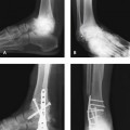
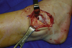
The triad of recurrent ankle sprains, heel varus, and stress fracture of the fifth metatarsal should always be taken into consideration when treatment is planned in the sedentary or athletic patient (Figure 31-1). The case illustrated in Figure 31-1 represents a good example of failure of treatment if the underlying biomechanical and anatomic process is ignored. A calcaneus osteotomy, in addition to a stronger ankle ligament repair, which might have prevented the subsequent problems, would have provided improved correction. An ankle ligament reconstruction can be performed without correcting the heel varus, but the outcome will depend on the flexibility of the subtalar joint and presence of additional symptoms.


If, for example, the patient has symptoms while walking on a flat ground surface and heel varus is present, then my inclination is to correct the calcaneus at the same time. If a patient has undergone previous ankle ligament reconstruction and has recurrent symptoms, I always look for unrecognized heel varus or mild tibia vara as a source for the failure. In most patients with heel varus, the deformity is bilateral. I have not, however, seen any biomechanical problem with correction of the hindfoot varus on one heel alone. Patients seem to adapt to this correction fairly well, and if contralateral symptoms develop subsequently, the correction can be done at a later date.


Usually an orthotic arch support with correct posting is sufficient to alleviate any minor symptoms of heel varus in the asymptomatic ankle. If a calcaneal osteotomy is necessary, it is performed in either one or two planes, depending on the pitch of the calcaneus. A lateral closing wedge osteotomy is always performed. Then the calcaneus can be translated slightly laterally and then shifted cephalad if the calcaneal pitch is markedly increased. For some patients with more severe heel varus associated with ankle instability, a plantar fascia release may need to be performed simultaneously.


The radiographic evaluation should routinely include weight-bearing radiographs, particularly if pain associated with the symptoms of instability is present. In addition to routine radiographs, I obtain MRI and CT scans as needed on the basis of additional disease present.


Assessment of the strength and function of the peroneal tendons in all patients who have recurrent ankle instability is important. Generally, these tendons are weak, and peroneal tendon rehabilitation is useful, even before the ankle ligament reconstruction is started. An appropriate rehabilitation regimen will facilitate functional recovery with a return to sports activities. I do not generally obtain an MRI study of the ankle even in patients who have peroneal tendon symptoms, because the incision is simply modified, as noted previously, and the peroneal tendons are inspected.


Operation Selection


Ankle ligament reconstruction in the high-performance athlete must be approached differently. The peroneal tendon should not be sacrificed as part of a reconstructive procedure, and even using a strip of the peroneus brevis tendon is not warranted in the high-performance athlete. In this context, high performance refers also to the gymnast, the ballet dancer, and other athletes for whom pivoting on the pointed foot is important. If a patient has gross ankle instability and an anatomic repair is not thought to be sufficient, then augmentation of this anatomic repair with a hamstring graft can be performed. Under these circumstances, my preference is to perform a percutaneous hamstring allograft procedure in these athletes and not to resort to an open procedure.


Although a hamstring autograft reconstruction has been popularized recently, this reconstruction should be avoided in athletes who run and in those who participate in ball and racket sports. This reconstruction is particularly relevant in the sprinting athlete, in whom terminal flexion torque will be compromised if the hamstring is sacrificed.


When should an anatomic repair be performed as opposed to a reconstruction? I am inclined to perform a percutaneous procedure using a hamstring allograft with biointerference screws whenever possible. The percutaneous procedure is very close to being an anatomic procedure, and although of course the graft is not inserted at the kinematic points on the fibula, this repair is an acceptable alternative in certain active and athletic patients. If a graft is not available, then either of two alternatives have to be considered: a modification of either the Elmslie or Chrisman-Snook procedure, or the hamstring procedure with an autograft. Although in the past I routinely used an anatomic repair of the Broström procedure for most reconstructions, for certain patients—the heavyweight athlete, such as a boxer or body builder, and the patient with any heel varus of any degree—who are not ideal candidates for this repair, I elected to use a tendon graft procedure. Whenever a tendon reconstruction procedure is performed, maintaining the correct kinematics of the ankle, which is impossible with use of a slip of the peroneal tendon, is important. Accordingly, a free tendon graft is preferable. Even with the allograft hamstring procedure, careful attention to selection of the entrance and exit points of the graft in the fibula is essential. As noted earlier, caution is indicated with the use of hamstring autograft in the sprinting athlete, which causes a deficit in terminal flexion torque.


In the patient with intraarticular ankle disease, the timing of the surgery is always a concern. For example, if an osteochondral defect requiring treatment is present, how should this operation be performed, in addition to ankle ligament reconstruction? (Figure 31-2). In patients with such defects, ankle arthroscopy, in conjunction with the ligament reconstruction, is recommended.


Traditional rehabilitation after ligament reconstruction, however, consists of immobilization for up to 6 weeks, presumably with considerable negative effects on recovery and rehabilitation after debridement for an osteochondral defect. Use of immobilization in this setting is essentially outmoded and should be discontinued except in rare instances. Although immobilization in a boot or brace can be used for comfort purposes, as a practical matter, if fixation techniques are used correctly, immobilization should not be needed at all. After any ankle ligament reconstruction, passive range-of-motion exercises are begun at 2 weeks, and the patient is permitted to walk out of the boot with a stirrup brace at 5 to 6 weeks, with physical therapy and rehabilitation started as early as possible.


Nonetheless, it is something to consider when combined diseases are addressed. The other issue pertaining to the intraarticular disease concerns the type of reconstructive procedure used. After ankle arthroscopy, interstitial tissue edema is always present, with fluid leakage into the soft tissues, and finding the correct anatomic plane—for example, for reconstruction using a Broström procedure—can be more difficult because of the tissue edema. I do not believe that performing an ankle arthroscopy is necessary in the absence of intraarticular disease or symptoms of ankle pain.


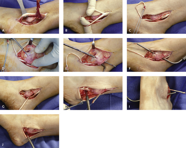
The Broström Procedure


I do not use the traditional “hockey stick”, or J-type, incision for the Broström procedure, because it does not permit visualization of the peroneal tendons. I use a more longitudinal incision located over the anterior fibula, inspecting the peroneal tendon simultaneously and facilitating repair of the calcaneal fibular ligament (Figure 31-3). This incision affords ready access to the anterior ankle and can even be extended to perform an open cheilectomy.


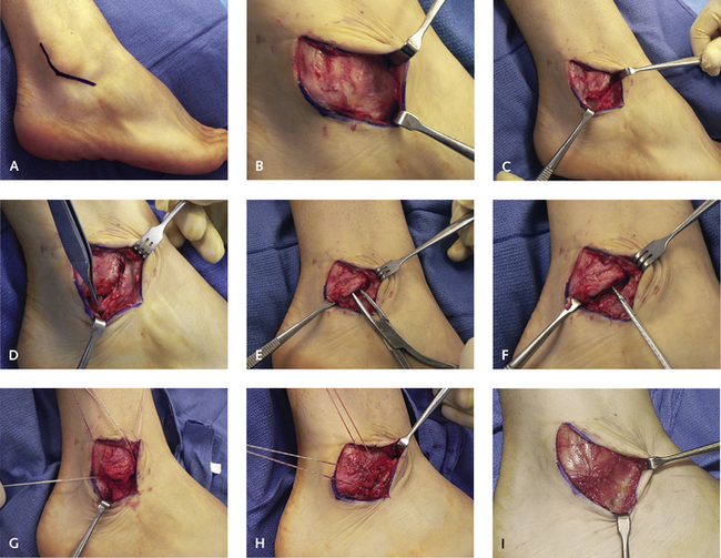
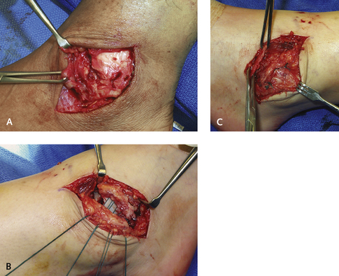
Care must be taken to avoid the superficial peroneal nerve anteriorly in the terminal portion of the incision. The soft tissue is reflected and the extensor retinaculum identified as a separate layer before the ankle joint is opened. This extensor retinaculum can be used to strengthen the anatomic repair (Figure 31-4). The inferior root of the extensor retinaculum inserts into the neck of the calcaneus just anterior to the subtalar joint, and can be used to stabilize both the ankle and subtalar joints in cases of combined instability.


The extensor retinaculum is not strong enough on its own to correct instability, and repair based on this structure should be combined with the anatomic procedure described. The incision through the anterior talofibular ligament must be made carefully. Sometimes a bony avulsion is present off the tip of the fibula; therefore the incision through the ligament must be as close to the fibula as possible. The original description of this procedure included a “vest over pants” repair of the anterior talofibular ligament, which is almost impossible to perform correctly because of the paucity of adequate ligamentous tissue. For this reason, I make the incision through the anterior talofibular ligament as close to the fibula as possible and dissect the ligament off the tip of the fibula. The periosteal tissue is then raised with a small cuff of the remnant of the anterior talofibular ligament off the fibula, and this can then be used to lie over the anatomic repair once the anterior talofibular ligament has been pulled up into the fibula. I generally debride the edge of the fibula, using either a rongeur or a small burr, to create a bleeding trough for reattachment of the ligament.


The same principle applies with use of the calcaneal fibular ligament. Detaching it directly from the fibula is easier than cutting it in its central body and then attempting a repair of a short ligament. Attachment of the anterior talofibular ligament to the fibula can be done either with a suture anchor or with sutures passed through Kirschner wire (K-wire) holes through the fibula. I prefer the latter technique because these holes can be made in pairs and the suture can be inserted through the ligament as a Y-shaped suture, pulled up, and imbricated with the ligament into the prepared trough on the tip of the fibula. Two sets of sutures are used to reattach the anterior talofibular ligament. When the ligament is tied down, the knot should not be placed over the fibula. The knot is invariably prominent, which can be irritating and painful, particularly in patients who have thin subcutaneous tissue. The knot should therefore always be tied on the ligament side of the repair, rather than on the bone. I use a nonabsorbable suture on a stout-tapered needle, passed through the predrilled holes in the fibula.


The peroneal tendons must be retracted completely to visualize the calcaneal fibular ligament. It is useful to prepare the sutures for both the anterior talofibular and calcaneofibular ligament before tying off the anterior talofibular ligament. Afterward, the ankle needs to be moved around, and the suture repair on the anterior talofibular ligament should not be disturbed when the calcaneofibular ligament is tied off. The anterior talofibular ligament is tied off first, with the foot in neutral dorsiflexion and slight eversion. Overtightening the ligaments with this technique is possible, and the foot should not be in dorsiflexion or forced eversion during the repair. At the completion of the repair of the anterior talofibular and calcaneofibular ligaments, the extensor retinaculum can be pulled up and sutured to the prepared flap of the periosteum and remnant of the anterior talofibular ligament over the fibula. Not all patients have a well-defined extensor retinaculum, and this part of the procedure is not always feasible. These final sutures must be buried; otherwise, they can serve as a source of irritation. I use absorbable sutures here for this reason, because even with a buried suture, the knot can be irritating.


Modification of the Chrisman-Snook Procedure


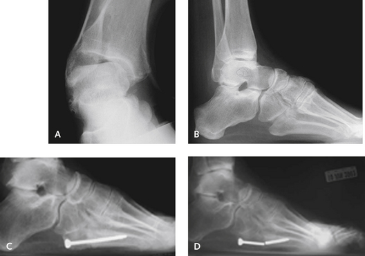
The original description of the Chrisman-Snook procedure included a long strip of the peroneus brevis tendon, which was split in half and placed through a bone tunnel in the calcaneus. This aspect of the technique is not necessary because a short strip of the anterior third of the peroneus brevis tendon is sufficient. The incision is made paralleling the peroneal tendons and extending for no more than 6 cm proximal to the tip of the fibula. The length of tendon that is required should be measured before the tendon is cut proximally, but rarely exceeds approximately 8 cm. The advantage of this procedure is that it can be used in the presence of a severe tear of the peroneus brevis tendon, for which a split portion of the tendon can be incorporated and used for the ligament reconstruction. Splits in the peroneus brevis tendon are common in combination with recurrent ankle instability, and if these splits are present, this portion of the tendon is then cut proximally and used for the reconstruction (Figure 31-5).


Mar 6, 2016 | Posted by in Reconstructive surgery | Comments Off on Ankle Instability and Impingement Syndromes

Full access? Get Clinical Tree

Get Clinical Tree app for offline access