(1)
Department of Neurosurgery Faculty of Medicine, Saga University, Saga, Japan
Keywords
Microvascular decompressionIts surgical anatomyCranial nerves and offending vesselsTheir imaging anatomyStitched sling retraction technique10.1 Introduction
To successfully perform microvascular decompression (MVD) procedures for neurovascular compression syndromes such as trigeminal neuralgia (TN), hemifacial spasm (HFS), and glossopharyngeal neuralgia (GPN), surgeons should be extremely familiar with the anatomical relationships between blood vessels and cranial nerves (CNs) in the cerebellopontine angle. The relationships between CNs and cerebellar arteries in the posterior cranial fossa are demonstrated in an autopsy specimen in Fig. 10.1a. Compression of CN V by the lateral pontomesencephalic segment of the superior cerebellar artery (SCA) is demonstrated in Fig. 10.1b.
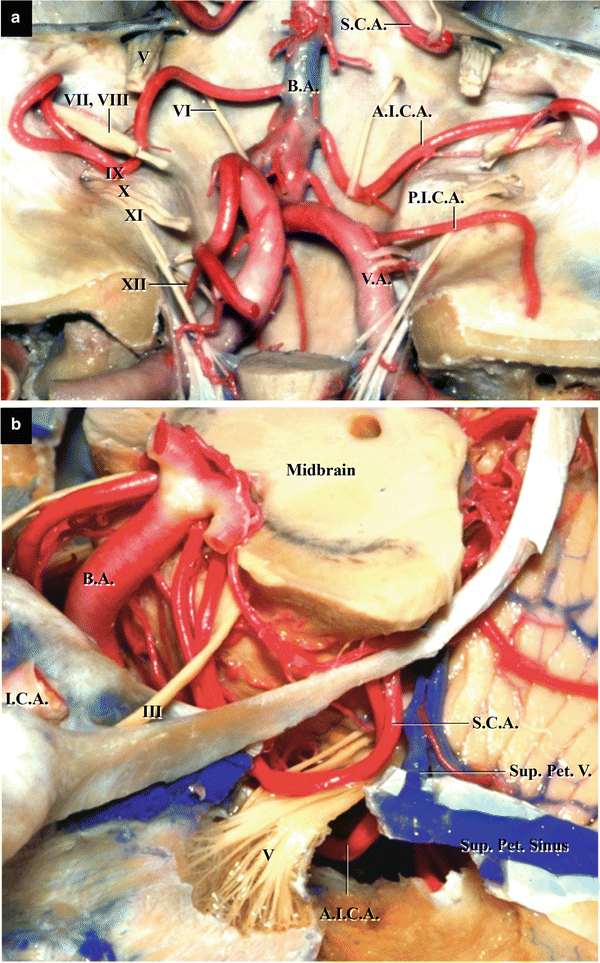

Fig. 10.1
Relationships between CNs and arteries in the posterior cranial fossa (from Matsushima T et al. [9] with permission). (a) The three cerebellar arteries and CNs; posterior view. (b) The lateral pontomesencephalic segment of the left SCA compressing the left trigeminal nerve (CN V). Left superolateral view
Preoperative magnetic resonance imaging (MRI) is used not only to identify offending vessels but also to estimate intraoperative findings and views. The basic anatomy of the three cerebellar arteries is described in Chap. 3: “Three Cerebellar Arteries: Superior Cerebellar Artery, Anterior Inferior Cerebellar Artery, and Posterior Inferior Cerebellar Artery.” In particular, this chapter explains their anatomical variations and relationships with CNs in detail and comments on their appearances on MRI. Furthermore, because we employ the stitched sling retraction technique as an MVD technique, we explain the surgical anatomy relevant to the technique. Subsequently, the approaches and techniques for the three MVDs will be independently discussed in Chaps. 11, 12, and 13. We utilize three different approaches in MVDs for TN, HFS, and GPN [3, 10, 12–14, 19].
10.2 Trigeminal Neuralgia
10.2.1 Offending Vessels
The offending vessels in TN include SCA, the anterior inferior cerebellar artery (AICA), vertebral artery (VA), and superior petrosal vein (Sup. Pet. V.) [2, 12, 16]. Multiple vessels are often responsible for TN. SCA is the most common offending vessel, but in a few cases, veins alone are responsible [16].
10.2.2 SCA and AICA and Their Relationships to CN V
SCA that originates from the distal portion of the basilar artery (BA) encircles the midbrain. When it runs between CNs IV and V, it compresses CN V from the superomedial side. Further, it passes through the cerebellomesencephalic fissure, the groove made by the midbrain, and the cerebellar hemisphere (Fig. 10.2a, b). SCA is divided into four segments: the anterior pontomesencephalic, lateral pontomesencephalic, cerebellomesencephalic, and cortical segments [1]. When the lateral pontomesencephalic segment bifurcates into the rostral and caudal trunks, it forms a downward loop to compress CN V from the superomedial side (Figs. 10.1b and 10.2a–c). Therefore, this artery may compress the nerve at its multiple sites depending on the point of its bifurcation and elongation of the downward loop [2, 12] (Fig. 10.3). When SCA bifurcates before arriving at CN V and forms a long downward loop, both the rostral and caudal trunks may compress CN V at a maximum of four points on SCA. On the other hand, when SCA bifurcates after passing CN V, it compresses the nerve at one or two points by its main trunk. In some cases, after passing the CN VII and VIII complex, AICA forms a long upward loop that compresses CN V from below. In such cases, CN V is sandwiched by SCA and AICA from both the superomedial and inferolateral sides (Figs. 10.2c and 10.3g). As it is difficult to observe the entry zone of CN V because of an obstacle of the cerebellar hemisphere through the operative view, we have to search the course of the caudal trunk of SCA preoperatively.
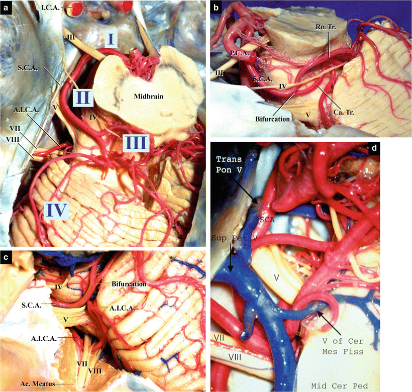
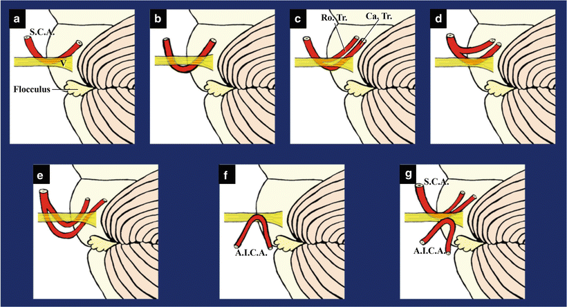

Fig. 10.2
CN V and surrounding vessels (from Matsushima T et al. [21] with permission). (a) The left SCA, its course and segmentation; superior view. (b) The left SCA, the lateral pontomesencephalic segment, and its bifurcation into the rostral and caudal trunks; lateral view. (c) The left CN V sandwiched by SCA and AICA; lateral view. (d) Veins surrounding the left CN V; superior view

Fig. 10.3
Schematic illustrations showing the various relationships between the SCA and/or AICA and the trigeminal nerve (CN V); lateral view (from Matsushima T et al. [21] with permission)
10.2.3 The Sup. Pet. V. and CN V
There are usually a few common stems of the Sup. Pet. V. that drain into the superior petrosal sinus near the Meckel’s cave, gathering tributaries from the anterolateral surface of the pons, posterior surface of the midbrain, and petrosal cerebellar hemisphere around the flocculus [9, 11, 22]. They often make a contact with CN V near the Meckel’s cave and sometimes cause TN. Offending veins in TN include the transverse pontine vein, which runs from the anterior aspect of the pons to the Meckel’s cave, and the vein of the cerebellopontine fissure, which runs over CNs VII and VIII from the flocculus [16] (Fig. 10.2d). In cases of TN, causative lesions may occur along all sites of CN V in the posterior cranial fossa, and all veins have the potential to become offending vessels. Thus, surgeons should avoid losing sight of the offending veins. In particular, in a case of the transverse pontine vein, which courses in the deep area and is often hidden by CN V, it is important to examine the deep area surrounding the Meckel’s cave thoroughly [16]. In many cases, arachnoiditis is also observed around CN V near the Meckel’s cave.
(Regarding the Sup. Pet. V., refer to Chap. 5: “The Bridging Veins in the Posterior Cranial Fossa.”)
10.2.4 CN V and Offending Vessels on Magnetic Resonance Imaging (MRI)
The aforementioned relationships between the offending vessels and CN V can be visualized on MRI and magnetic resonance angiography (MRA). Usually, original MRA images and/or axial views of T2 reversed-weighted images can be used to observe the relationships between CN V and surrounding vessels. Cross sections of SCA are usually observed medial to CN V and Sup. Pet. V. lateral to the nerve (Fig. 10.4a, b). Sagittal view of MRI reveals SCA making a downward loop and compressing the nerve (Fig. 10.4b). Three-dimensional (3D) images are helpful for easy understanding of these relationships and preoperative anticipation of an operative view (Fig. 10.4c, d).
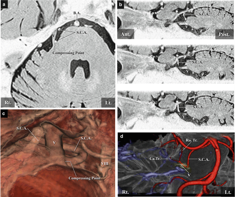

Fig. 10.4
Preoperative images in cases of right trigeminal neuralgia. (a) T2-reversed-weighted image; axial view. (b) T2-reversed-weighted image; sagittal view. (c) 3D image showing the intraoperative findings, the downward loop of the right SCA compressing the right CN V from the medial side; lateral view. (d) 3D image showing the right SCA, its course, bifurcation, and relationship to CN V; right anterolateral view
10.3 Hemifacial Spasm
10.3.1 Offending Vessels
The offending vessels in HFS are arteries such as AICA, posterior inferior cerebellar artery (PICA), and/or VA. It is safe to say that veins are never offending vessels in HFS. HFS caused by vascular compression syndrome usually occurs at the exit zone of CN VII in the supraolivary fossette; when the arteries change their course in this region, they compress the exit zone of the nerve and cause HFS. In many cases, there are multiple offending arteries [15, 17]. AICA often entwines nerve bundles of CNs VII and VIII when passing in front of the porus acusticus, forming a neurovascular complex (Fig. 10.5b). However, this distal portion is not responsible for HFS [15, 18].
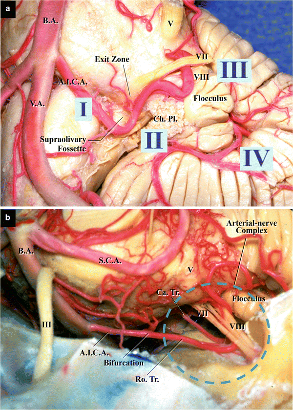

Fig. 10.5
The facial nerve (CN VII) and AICA (from Matsushima T et al. [21] with permission). (a) The left AICA, its course, and segmentation; anterior view. (b) The left AICA and its bifurcation into the rostral and caudal trunks; left superolateral view
10.3.2 AICA, PICA, and Their Relationships with the Exit Zone of CN VII
After originating from the proximal portion of BA, AICA runs laterally near the pontomedullary sulcus and changes its direction at the supraolivary fossette, heading toward the porus acusticus internus. It travels forward to the auditory canal, parallel to the nerve bundles of CNs VII and VIII (Fig. 10.5a, b


Stay updated, free articles. Join our Telegram channel

Full access? Get Clinical Tree


