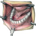
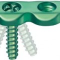
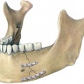
31 Alveolar Bone Distraction Diminished vertical height of the alveolar ridge often leads to loss of vertical dimension of the face, which causes impaired masticatory function and poor facial aesthetics. The alveolar ridge reduction may be caused by traumatic injury, tumor resection, and bone destruction with periodontosis, or general atrophy accompanying tooth loss or old age. In many of these cases, dentures cannot be worn due to lack of mechanical retention. Alternatively, dental implants have been inserted in such situations, where a sufficient amount of bone is available. In order for dentoalveolar implants to be successful, however, a minimum height and width of bone is required. Where too little bone is available, the alveolar ridge is often augmented using techniques such as guided bone regeneration, onlay grafts, or implantation of alloplastic material. Bone grafts, however, involve an increased surgical risk and consequently a high complication rate. Donor site morbidity and considerable resorption of the grafted bone occur within the year following the procedure (Chiapasco, Zaniboni, and Rimondini, 2007). In light of these problems, several groups have been developing new techniques of vertical alveolar ridge augmentation based on the principles of callus distraction established by the Russian orthopedic surgeon, Gavriel Ilizarov (1989a, b), who pioneered the reconstruction technique of distraction osteogenesis for managing a variety of limb deformities. The initial work on alveolar ridge distraction was done in dog experiments by Block, Chang, and Crawford (1996), who demonstrated histological evidence of regenerated bone formation during alveolar ridge distraction. Since 1996 our medical team at the University of Cologne has also been working on alveolar ridge augmentation with the vertical distraction osteogenesis technique. Using the Ilizarov protocol as a basis, we have applied this technique for vertical augmentation of both the dentulous and edentulous alveolar process. Fig. 31.1 Osteotomy of the segmental cranial part of the toothless bone. The distractor has been inserted. In edentulous parts of the mandible after segmental re-section in tumor surgery or trauma loss, reconstruction by vertical distraction can be used instead of bone transplantation (Fig. 31.1). After osteotomy the segmented cranial part of the toothless bony gap of the mandible is mobilized. The vascular supply of this segment is dependent on the preservation of the alveolar attachment and the lingual or the palatal mucoperiosteal flap. Afterward the distractor can be inserted (see Fig. 31.1) and the bony segment is stabilized for 7 days. The regimen for callus distraction is 0.5 mm, twice each day. In compromising tissues with scaring or after radiotherapy less activation is recommended (Amir et al., 2006). Within 10 days a previously estimated vertical increase of 10 mm can be obtained. Four weeks later, mineralization of the new bone in the distracted gap can be seen to be starting (Fig. 31.2), as well as reunion of the transported bone in the alveolar ridge. Early mineralization studies suggest that insertion of dental implants is possible after 3 months (Yamamoto et al., 1997).
Introduction

Technique
Related posts:

Stay updated, free articles. Join our Telegram channel

Full access? Get Clinical Tree


