Classification
Depth
Examples
Very superficial-light peels
Necrosis up to the level of stratum corneum
TCA 10%, Glycolic Acid (GA) 30–50%, Salicylic acid 20–30%, Jessner’s solution (1–3 coats), Tretinoin 1–5%
Superficial-light peels
Necrosis through the entire epidermis up to basal layer
TCA 10–30%, GA 50–70%, Jessner’s solution (4–7 coats)
Medium-depth peel s
Necrosis up to upper reticular dermis
TCA 35–50%, GA 70% plus TCA 35%, 88% phenol un-occluded, Jessner’s solution plus TCA 35%, solid CO2 plus TCA 35%
Deep peels
Necrosis up to mid-reticular dermis
Baker-Gordon phenol peel
Indications
The preprocedure consultation is essential to identify patients who are ideal candidates for intervention as well as to identify at-risk patients who are best avoided or who require an extra-cautious approach. The current indications for medium-depth peels include epidermal lesions , pigmentary disorders , acne issues, and aesthetic (Table 2) (Monheit and Kayal 2003; Khunger 2008; Rendon et al. 2010; Patel et al. 2014; Hession and Graber 2015). The mild depth can also be used in blending of the effects of deeper resurfacing procedures (Bolognia et al. 2012; Tung and Rubin 2011).
Table 2
Possible indications of medium-depth chemical peels
Epidermal lesions |
Seborrheic keratoses Actinic keratoses Warts Milia Sebaceous hyperplasia Dermatoses papulosa nigra |
Pigmentary disorders |
Melasma Postinflammatory hyperpigmentation Freckles Lentigines Facial melanoses |
Acne related |
Superficial to mild acne scars Postacne pigmentation Comedonal acne Acne excoriee Acne vulgaris-mild to moderately severe acne |
Aesthetic |
Photoaging Fine superficial wrinkling Dilated pores Superficial scars |
Photoaging is one of the conditions for which skin mild-depth peels are most often performed and in these patients, the Glogau scale (Table 3) is very helpful in the peel depth choice decision (Glogau 1996). Patients in type I can be managed with superficial chemical peels or microdermabrasion in association with medical cosmeceutical therapy (e.g., glycolic acids, topical retinoids, active cosmeceutical formulas) as they are frequently youthful with minimal to mild photoaging. Individuals in type II are better treated with a medium-depth chemical peel associated with a long-term medical therapy including an α-hydroxy acid (AHA) and/or a retinoid. Patients in category III normally need prolonged medical treatment with a medium-depth chemical peel (with or without dermasanding), a deep chemical peel, dermabrasion, laser resurfacing or associations between them. In type IV patients, the therapies described to type III would certainly be indicated; however, invasive surgical operation such as blepharoplasty, rhytidectomy, scar revision, and others are frequently needed in addition to achieve the expected results (Bolognia et al. 2012; Tung and Rubin 2011; Khunger 2008; Monheit 2004).
Table 3
Glogau photoaging classification
Type I – No wrinkles |
Early photoaging: mild pigmentary disorders, minimal wrinkles, no keratosis Patients typical age (years): 20s or 30s Makeup: Minimal or none |
Type II – Wrinkles in motion |
Early to moderate photoaging: Early senil lentigines visible, parallel smile lines beginning to appear, keratosis palpable but not visible Patients typical age (years): 30s or 40s Makeup: Usually wear some foundation |
Type III – Wrinkles at rest |
Advanced photoaging: Obvious dyschromia, telangiectasias, visible keratosis, wrinkles even when not moving facial muscles Patients typical age (years): 50s or older Makeup: Wears heavy foundation |
Type IV – Only wrinkles |
Severe photoaging: yellow-gray color of skin, prior skin malignancies, wrinkled throughout with no normal skin Patients typical age (years): 60–70s Makeup: Cannot wear (“cakes and cracks”) |
During the discussion on correct indication, some data are of extreme relevance. Head and neck are the most important areas due to their aesthetic value, and one must take caution when treating the neck due to its propensity for complications. It is known that areas with more pilosebaceous units have better re-epithelialization (Bolognia et al. 2012). Also beware when indicating procedure to hands and arms because they have less predictable and less impressive results (Gadelha and Costa 2009). Lesions originated in the epidermis (actinic keratosis, lentigines) have betters responses to chemical peels than lesions originated in the dermis (Tosti et al. 2006).
The indication also depends on the patient’s tolerances and expectations for correcting his skin condition. Some individuals do not wish to enhance skin surface regardless of serious issues, and others desire marked improvement in relatively minor problem areas. The condition severity and the patient wishes will lead the treatment (Gadelha and Costa 2009). These wishes ought to be tempered with data on what is conceivable and what is alluring for the patient regarding treatment. Approach every patient honestly, discussing about plausible outcomes, risks , advantages, and alternatives (Gadelha and Costa 2009; Bolognia et al. 2012).
Contraindications
History of AIDS, hepatitis, immunosuppressive systemic diseases, or usage of immunosuppressive medication must be identified, as they seem to grant higher frequency of secondary infection after the procedure. In a similar way, a history of abnormal scars or keloids deserves more attention as these patients may end up with an unpleasant outcome. Fitzpatrick sun-reactive skin type classification is another concern, as skin types IV, V, and VI tend to develop postinflammatory hyperpigmentation (Table 4) (Rullan and Karam 2010; Monheit 1995). Patients in use of contraceptives, supplemental hormones, or minocycline should be alerted due to the high risk of postinflammatory hyperpigmentation. Always question about history of facial surgery, prior resurfacing procedure, or oral isotretinoin use in the last 6 months as these can also increase complications (Dingman et al. 1994; Rubenstein et al. 1986). History of radiation therapy is important as it can destroy the pilosebaceous units, which are essential to re-epithelialization (Wolfe 1982).
Table 4
Fitzpatrick’s classification of sun-reactive skin types
Skin type | Color | Reaction to first summer exposure |
|---|---|---|
I | White | Always burn, never tan |
II | White | Usually burn, tan with difficulty |
III | White | Sometimes mild burn, tan average |
IV | Moderate brown | Rarely burn, tan with ease |
V | Dark browna | Very rarely burn, tan very easily |
VI | Black | No burn, tan very easily |
Special care should be taken in patients with some dermatologic diseases. Vitiligo and psoriasis can be exacerbated due to the isomorphic response. The vasomotor instability in rosacea can lead to an exaggerated inflammatory response postprocedure (Gadelha and Costa 2009). Connective autoimmune diseases, such as cutaneous lupus and scleroderma, can be activated by the chemical peeling trauma. The contraindications in mild-depth peels can be divided into absolute and relative (Table 5).
Table 5
Contraindications to mild-depth and deep peels
Absolute |
Open wounds Active infections (bacterial, viral, or fungal) Use of oral isotretinoin in the last 6 months Pregnancy History of drugs with photosensitizing potential Patient with unrealistic expectations Uncooperative patient (patient is careless about sun exposure or application of medications) Poor physician-patient relationship |
Relative |
Recent facial surgery in the last 6 months History of abnormal scar formation or delayed wound healing History of hyperpigmentation History of therapeutic radiation exposure Fitzpatrick Phototype IV, V, and VI History of active dermatologic diseases such as seborrheic dermatitis, rosacea, atopic dermatitis, vitiligo, contact dermatitis, and psoriasis |
Besides the contraindications, it is also important to consider the presence of inflammation (seborrhea, retinoid dermatitis, etc.) and skin translucency as the more inflammation present and the more translucent the skin is, the more likely the peeling will increase its depth, leading to possible complications. Have in mind that telangiectasia treatment is not a good chemical peeling indication because it has an unsatisfactory response. Besides, the peeling can also exacerbate them by removing the pigmentary irregularities (Bolognia et al. 2012; Tung and Rubin 2011).
Patients must completely understand the peeling limitations, pre and post care, risks and potential benefits, and must sign an informed consent prior to the procedure. If you feel vulnerability in physician-patient relationship, do not perform the procedure.
Trichloroacetic Acid Peel
Trichloroacetic acid (Fig. 1) is very versatile as it can be used to create very superficial, superficial and medium-depth peels depending on the concentration of TCA used (Table 1), adequacy of skin priming, coats of acid applied, and technique of application. TCA is most commonly used for medium-depth peels, especially to treat pigmentation disorders and early facial rhytides (Lee et al. 2002).
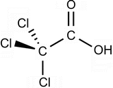
Fig. 1
Molecular structure of TCA
TCA is a caustic substance obtained through distillation of the product from nitric acid steam on chloral acid. It is found in our environment as an herbicide, as a major metabolite of dry cleaning process and as chemical peels. It has almost nonexistent toxicity, even when applied in concentrated form on the skin. TCA has the lowest pKa and is stronger than any other acids used as chemical peels. As it progresses through the distinctive skin layers, the acid peeling is “neutralized,” prompting a coagulation of skin proteins.
Its action is proportional to the concentration and to the amount applied. Higher concentrations of TCA lead to more acidic solutions and deeper penetrations. Higher number of coats or more pressure applied during the procedure also lead to deeper penetration. Note that depending on the skin area, higher number of coats needs to be applied (at same concentration) to achieve the same level of frosting (Fig. 2). As a peculiar characteristic of TCA, its visual changes (from light speckling to white frost) following application in the skin indicate degree of protein coagulation (Tung and Rubin 2011; Landau 2008).
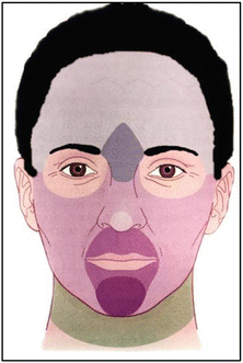
Fig. 2


Skin reactivity to TCA coating. Higher number of coats needs to be applied in darker areas (at same concentration) to achieve the same level of frosting
Related posts:

Stay updated, free articles. Join our Telegram channel
Full access? Get Clinical Tree
