Chapter 1 Abdominal Wall Anatomy and Vascular Supply
1 Clinical Anatomy
1 Overview
 The anterior abdominal wall (Figs. 1-1 to 1-3) is a hexagonal area defined superiorly by the costal margin and xiphoid process; laterally by the midaxillary line; and inferiorly by the symphysis pubis, pubic tubercle, inguinal ligament, anterior superior iliac spine, and iliac crest.
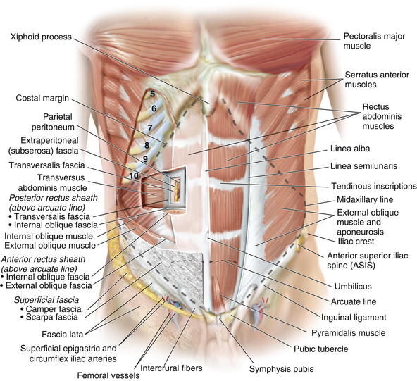

2 Superficial Fascial Layers (see Figs. 1-1 and 1-2)
 The superficial fascia of the abdominal wall consists of a single layer above the umbilicus, consisting of the fused Camper and Scarpa fasciae.

 Scarpa fascia fuses inferiorly with the fascia lata of the thigh and continues posteriorly to the perineum, where it is called Colles fascia.
3 Deep Fascial Layers (see Figs. 1-1 and 1-2)


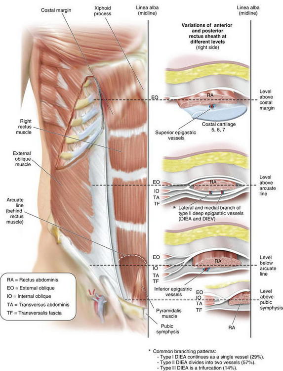
 The arcuate line (see Fig. 1-3) is located midway between the umbilicus and symphysis pubis and is a transition point where the posterior rectus sheath transitions from being the fusion of part of internal oblique fascia and transversalis fascia superiorly to only transversalis fascia inferiorly.
 Below the arcuate line, the external oblique and internal oblique fasciae merge to form the anterior rectus sheath. The posterior rectus sheath consists of transversus abdominis fascia, making this only a thin layer with minimal strength.
 The linea alba results from fusion of the anterior and posterior rectus sheaths and lies in the midline, extending cranially from the xiphoid process to the pubic symphysis caudally Figure 1-4 shows the anterior wall fascia after dissection of the abdominal wall skin and subcutaneous tissue, showing the linea alba and linea semilunaris.
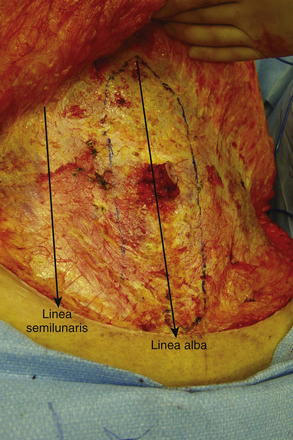
 Pearls and Pitfalls
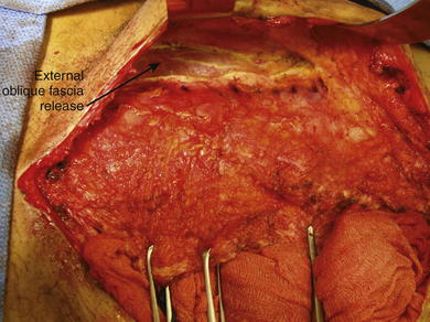
Incision, release, and dissection of the anterior external oblique fascia can be done for repair of ventral hernias. This technique is called the components separation (Fig. 1-5). The incision in the external oblique fascia is made 1 to 2 cm lateral to the linea semilunaris, and the fascia is released to attain primary closure. Incisions also can be made in the posterior rectus sheath to gain additional length.
4 Abdominal Wall Musculature (see Figs. 1-1 to 1-3)
 The paired rectus abdominis muscles are the principal flexors of the anterior abdominal wall. They function to stabilize the pelvis while walking. They also protect the abdominal organs and help in forced expiration.
 The rectus abdominis muscles originate from the pubic symphysis and pubic crest and insert on the anterior surfaces of the fifth, sixth, and seventh costal cartilages and the xiphoid processes. Laterally, the rectus sheath merges with the aponeurosis of the external oblique muscles to form the linea semilunaris (Fig. 1-4).
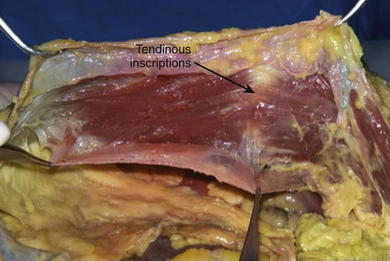
 Three to four tendinous inscriptions, which are adherent to the anterior rectus sheath, interrupt the rectus abdominis along its length (Fig. 1-6).

 The transversus abdominis muscle is the deepest of the three lateral abdominal wall muscles and courses in a horizontal direction. It originates from the anterior three fourths of the iliac crest; lateral third of the inguinal ligament; and inner surface of the lower six costal cartilages, interdigitating with fibers of the diaphragm. The muscle ends medially in a broad flat aponeurosis, merging above the arcuate line with the posterior lamella of the internal oblique aponeurosis and the linea alba. Below the arcuate line, it inserts into the pubic crest and pectineal line, forming the conjoint tendon with the internal oblique.


5 Neurovascular Supply of the Abdominal Wall
 Pearls and Pitfalls

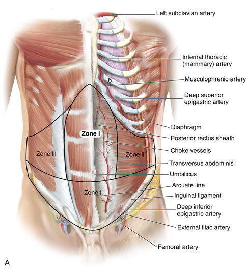
 Zone I consists of the upper and midcentral abdominal walls and is supplied by the vertically oriented deep superior (Fig. 1-7, A).and deep inferior epigastric arteries (Fig. 1-7, B).
 Zone II consists of the lower abdominal wall and is supplied by the epigastric arcade, superficial inferior epigastric, superficial external pudendal, and superficial circumflex iliac arteries. Perforators from the deep circumflex iliac arteries also supply a region of skin posterior and cephalad to the anterior superior iliac spine along the axis of the iliac crest.
 Zone III consists of the lateral abdominal wall (flank region) and is supplied by the musculophrenic, lower intercostals (Fig. 1-8, A), and lumbar arteries (Fig. 1-8, B).
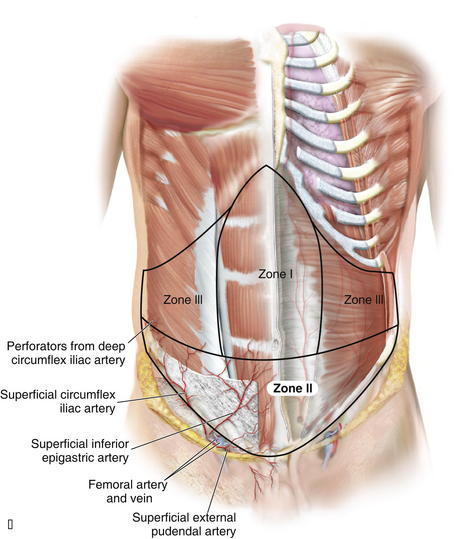
Stay updated, free articles. Join our Telegram channel

Full access? Get Clinical Tree


