Figure 2.1
Merkel cell carcinoma
Clinical Differential Diagnosis
(Raised erythematous lesion on sun exposed skin)
Squamous cell carcinoma
Actinic keratosis
Merkel cell carcinoma
Lymphoma
Angiosarcoma
Metastasis
Histopathology
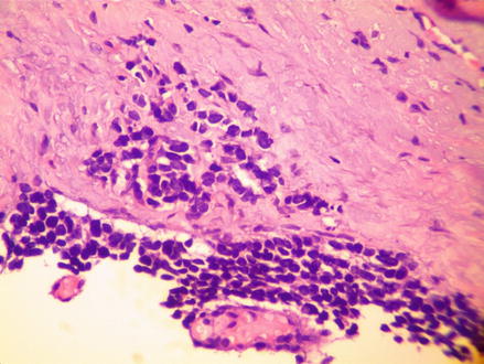
The lesion on the right cheek showed atypical nests of keratinocytes (Squamous cell carcinoma) while the left temple showed basal nests of various sizes (Basal cell carcinoma). The left forehead showed two distinct diseases. One was full thickness squamous atypia with overlying parakeratosis. The second showed irregular shaped sheets of small, round cells with scant amounts of cytoplasm (Fig. 2.2). Some areas of the tumor was dyscohesive resulting in scattered single cells in the dermis (Fig. 2.3). The nucleus showed a coarse granular (salt and pepper) pattern. These cells stained positive for cytokeratin 20 as perinuclear spots, but negative for MART (Fig. 2.4).
 6 Year Old White Male with Recurrent Brownish Patches Around Mouth
 37 Year Old White Male with Multiple, Brown, Purple Nodules on Arms and Legs
 70 Year Old White Male with Grouped Blisters on Right Lower Leg
 7 Year Old Male with an Enlarging, Round Area of Hair Loss
 29 Year Old White Female with Grouped Blisters on Left Thigh
 46 Year Old Female with Multiple Slightly Eroded Reddish Papules on Her Arm


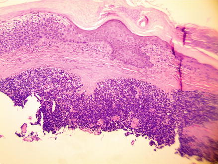
Figure 2.2
H&E 40×, Merkel cell carcinoma with overlying squamous cell carcinoma in situ on the left side of the epidermis
Related posts:
6 Year Old White Male with Recurrent Brownish Patches Around Mouth
37 Year Old White Male with Multiple, Brown, Purple Nodules on Arms and Legs
70 Year Old White Male with Grouped Blisters on Right Lower Leg
7 Year Old Male with an Enlarging, Round Area of Hair Loss
29 Year Old White Female with Grouped Blisters on Left Thigh
46 Year Old Female with Multiple Slightly Eroded Reddish Papules on Her Arm
Stay updated, free articles. Join our Telegram channel
Full access? Get Clinical Tree
