Procedure 93 Digital Ray Amputation
![]() See Video 69: Digital Ray Amputation
See Video 69: Digital Ray Amputation
Examination/Imaging
Clinical Examination
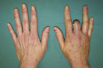
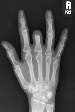
 In patients undergoing ray amputation for a painful stump, the point of maximal tenderness due to a neuroma (if any) should be identified preoperatively and marked. Figure 93-1 shows a patient with a painful stump.
Surgical Anatomy
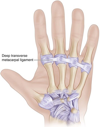
 The deep transverse metacarpal ligament connects the volar plates of the MCP joints of the index, long, ring, and small fingers. The flexor tendons, neurovascular bundles, and lumbrical tendon pass palmar to the deep transverse MCP ligament, whereas the interosseous tendon lies dorsal to it (Fig. 93-3).
Exposures
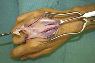
 A racket-shaped incision is marked around the base of the finger. On the dorsum, this incision extends over the proximal metacarpal in a longitudinal fashion (Fig. 93-4A). On the palmar aspect, it is designed as a V (Fig. 93-4B).


