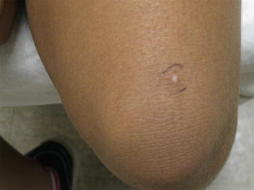
Figure 9.1
Verruca vulgaris

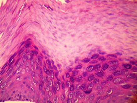
Figure 9.2
Verruca vulgaris
Clinical Differential Diagnosis
Epidermal nevus
Actinic keratosis
Seborrheic keratosis
Verruca vulgaris
Histopathology
The shave biopsy of the left knee was performed and measured 0.3 × 0.2 × 0.1 cm. The left posterior upper arm exhibited slight hyperkeratosis and papillomatosis with acanthosis. Within the epidermis, some keratinocytes exhibited a perinuclear vacuolization with irregular nuclear contours while other keratinocytes exhibited hypergranulotosis. A shave biopsy of the left posterior upper arm was performed and measured 0.3 × 0.2 × 0.1 cm (Figs. 9.1, 9.3, 9.4, and 9.5).


