Procedure 9 Staged Flexor Tendon Reconstruction
 See Video 6: Staged Flexor Tendon Reconstruction
Indications
 Finger that is not amenable to primary or delayed primary repair or a single-stage tendon graft procedure, as in the following cases:


Examination/Imaging
Clinical Examination
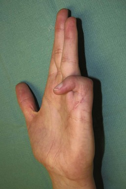
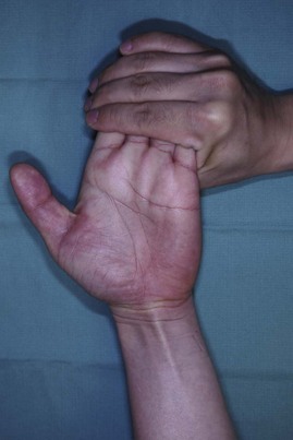
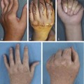
 The patient should be examined to identify specific tendon involvement and reconstructive needs. Patients will present with loss of active distal interphalangeal (DIP) and proximal interphalangeal (PIP) joint flexion if both the FDP and FDS are divided, or loss of only DIP joint flexion if only FDP has been injured. On inspection, the normal finger cascade is lost, with the affected digit in an extended position (see Fig. 9-2).
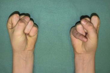
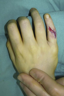
 The metacarpophalangeal (MCP) and interphalangeal (IP) joints should have full passive range of motion, or they will require capsulotomy before tendon reconstruction (Figs. 9-3 and 9-4).


Surgical Anatomy
 The lumbrical muscle prevents further proximal retraction of the FDP tendons. For this reason, the proximal juncture of the tendon graft is usually placed in the palm to the FDP, just distal to the lumbrical origin. The median innervated first and second lumbricals are unipennate and arise from the radial and palmar surface of the FDP tendons to the index and long finger. The ulnar innervated third and fourth lumbricals are bipennate and arise from contiguous surfaces of the long/ring finger FDP tendon and ring/small finger FDP tendon, respectively. If the palm is involved in trauma, the proximal juncture is placed in the forearm.


Stage I: Exploration, Pulley Reconstruction, and Placement of Silicone Rod
Exposures
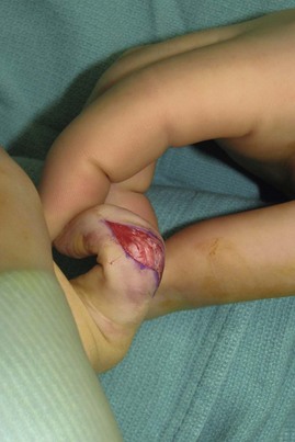
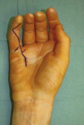
 Bruner incisions are designed from the mid-distal phalanx to the distal palm. Incisions are planned such that they incorporate any previous scars (Fig. 9-6).
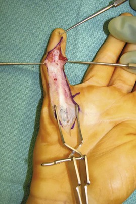
 Thick skin flaps are raised in a plane superficial to the tendon sheath to expose the tendon sheath from the A1 pulley to the DIP joint (Fig. 9-7).
Pearls
If pulley reconstruction is not necessary, two separate incisions may be used: the first for distal palm exposure and the second for exposure of the tendon sheath in the finger.
The neurovascular bundles should be identified outside the zone of injury (related to previous scars) and protected when flaps are raised, as well as for the remainder of the operation.
Procedure
Step 1: Assessment of Pulleys
 The tendon sheath is dissected carefully in an attempt to preserve any remnants of the A2 and A4 pulleys. Usually there is moderate to severe scarring, making it impossible to preserve good-quality pulleys (Fig. 9-8).
Step 2: Débridement of Scar Sheath and Tendons
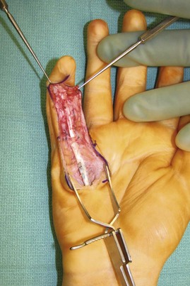
 The remnants of the scarred FDS and FDP tendons are excised distal to the lumbrical origin. Approximately 1 cm of the distal FDP insertion (if available) is preserved to provide a distal anchor point for the silicone rod.


Step 3: Pulley Reconstruction
 We use excised remnants of the FDS and FDP harvested from the finger or from FDS tendon in the palm or forearm to reconstruct the pulley.
Related posts:
 28: Pronator Teres Rerouting
 33: Correction of Swan-Neck Deformity in the Rheumatoid Hand
Stay updated, free articles. Join our Telegram channel

Full access? Get Clinical Tree


