Procedure 89 Total Wrist Fusion
![]() See Video 67: Total Wrist Fusion
See Video 67: Total Wrist Fusion
Indications
Exposures
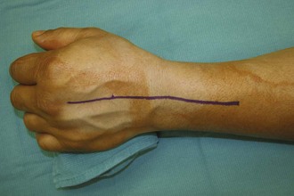
 A dorsal midline incision is used, centered at the radiocarpal joint and extending past the midpoint of the long finger metacarpal and proximally, about 5 cm proximal to the Lister tubercle (Fig. 89-2).
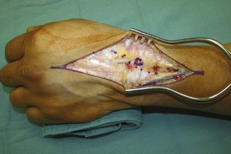
 Flaps are elevated, keeping cutaneous nerves in the flaps (Fig. 89-3).
 Incise third dorsal compartment and retract the extensor pollicis longus (EPL) tendon (Fig. 89-4).
 Raise retinacular flaps, exposing the second to fifth extensor compartments.
 The posterior interosseous nerve (PIN) is located on the floor of the fourth extensor compartment. A PIN excision is performed after retracting the extensor tendons.
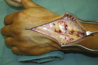
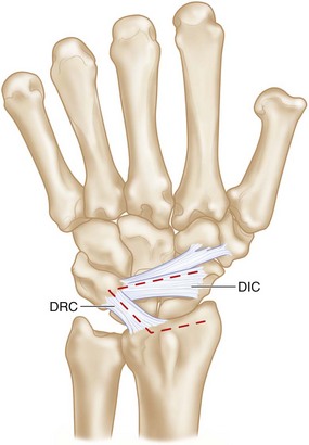
 A radially based capsular flap is formed by incising the dorsal intercarpal and dorsal radiocarpal ligaments and elevating them off the dorsal triquetrum (Fig. 89-5).
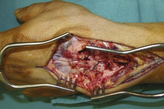
 Subperiosteal dissection is used to expose the dorsal radius, the carpus, and the long finger metacarpal (Fig. 89-6).


