Procedure 87 Four-Corner Fusion
![]() See Video 65: 4-Corner Fusion Using Kirschner Wires
See Video 65: 4-Corner Fusion Using Kirschner Wires
Examination/Imaging
Imaging
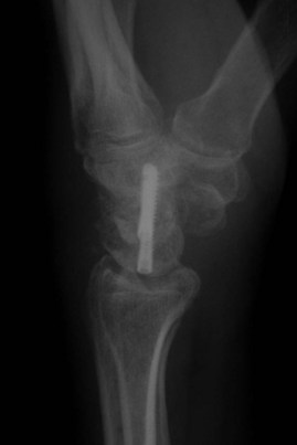
 Plain radiographs are usually sufficient for diagnosis. SLAC wrist, stage 3, demonstrates abnormal position of the scaphoid, scapholunate widening, radioscaphoid degenerative change and midcarpal arthrosis (Fig. 87-1).
 In cases in which there is uncertainty about the status of the radiolunate joint, advanced imaging (computed tomography) may be used.
Surgical Anatomy
 In either SLAC or SNAC, there will be widening at the scapholunate interval.
 Significant scaphoid flexion, lunate extension, and dorsal lunate prominence develop.
 Radioscaphoid degenerative change with styloid prominence and osteophytes is noted.
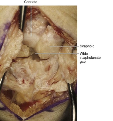
 Proximal scaphoid pole necrosis and/or arthrosis is present (Fig. 87-2).
 Scaphotrapeziotrapezoid arthrosis may be present.
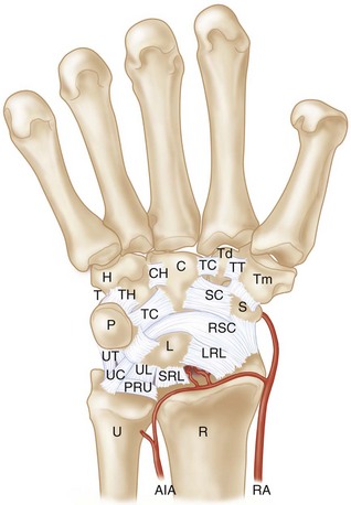
 The dorsal ligaments of the wrist—dorsal radiocarpal (DRC) and dorsal intercarpal (DIC)—have a conjoined insertion on the triquetrum. They provide the capsulotomy landmarks for wrist exposure.
 The volar extrinsic ligaments (radioscaphocapitate, short and long radiolunate) originate from the volar radius and extend obliquely to the carpus. These should be respected during scaphoid excision (Fig. 87-3).
Exposures
 A dorsal midline incision centered over the radiocarpal joint is used.
 Thick flaps are elevated dorsal to the retinaculum, keeping cutaneous nerves within these flaps.
 The retinaculum over the third dorsal compartment is incised.
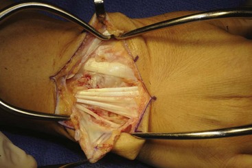
 Retinacular flaps are raised, exposing the second through fifth compartments (Fig. 87-4).
 The extensor tendons are retracted using retractors placed between the second and fourth compartments.
 A posterior interosseous neurectomy is performed.
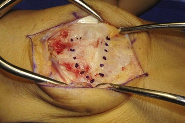
 A capsulotomy is performed by dividing the dorsal capsule along the dorsal intercarpal and dorsal radiocarpal ligaments (Fig. 87-5). The flap is elevated off the dorsum of the triquetrum and reflected, keeping it radially based (Fig. 87-6).
Stay updated, free articles. Join our Telegram channel

Full access? Get Clinical Tree


