Procedure 83 Darrach Procedure
![]() See Video 62: Darrach Procedure
See Video 62: Darrach Procedure
Indications
 Older, sedentary patients with posttraumatic distal radioulnar (DRU) joint arthritis, rheumatoid arthritis, and osteoarthritis without preexisting radiocarpal instability.
 Destruction of the triangular fibrocartilage complex with caput ulna syndrome and painful pronation and supination of the DRU joint.
 The Darrach procedure (distal ulna head excision) is preferred in patients with no ulnar translation of the carpus and low chance of translation in the future.
 In the setting of radiocarpal instability, the Darrach procedure can be performed concomitantly with a partial or total wrist fusion.
Examination/Imaging
Clinical Examination
 Evaluate active and passive wrist and DRU joint motion and compare with the opposite side. The affected wrist should be taken through full wrist motions of flexion-extension, pronation-supination, and radioulnar deviation with axial loading to assess stability and pain.
 Painful crepitus with pronation and supination indicate DRU joint arthritis that can be accentuated with manual compression of the joint.
 Dorsal wrist swelling can indicate extensor tendon synovitis. Palpation of the inflamed wrist is painful, and wrist mobility may be limited.
 Caput ulna syndrome is diagnosed as dorsal prominence of the ulnar head with accompanying ulnopalmar subluxation and supination of the carpus. The DRU joint will be swollen and painful on palpation. There may be painful limitation of pronation and supination.
 ECU subluxation and tendonitis should be distinguished from DRU joint arthritis by noting ECU subluxation with supination and ulnar deviation.
 Lunotriquetral (LT) instability from ligamentous tears should be distinguished from DRU joint arthritis by performing the LT shear test. The lunate is stabilized between the examiner’s thumb and index finger, and the triquetrum is sheared against the lunate in a dorsopalmar direction with the examiner’s other hand.
 The prominent caput ulna can cause extensor tendon ruptures, primarily at the fourth and fifth extensor compartments.
Imaging
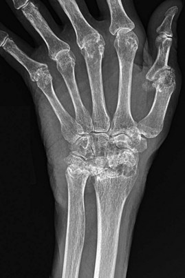
 Standard three views of the wrist are useful to assess extent of arthritis and wrist stability. Changes associated with arthritis include osteophytes, joint space narrowing, erosive changes, cystic lesions, and disuse osteopenia (Fig. 83-1).
 For differentiation of extensor tendon from joint synovitis, ultrasonography is the diagnostic tool of choice.
 Computed tomography and magnetic resonance imaging are typically not necessary in the evaluation of the DRU joint in rheumatoid arthritis.
Surgical Anatomy
 The dorsal sensory branch of the ulnar nerve lies in the subcutaneous tissue of the dorsal ulnar wrist.
 In the presence of caput ulna syndrome, the sixth dorsal compartment with the ECU is subluxed palmarly. The fifth dorsal compartment with the extensor digiti minimi (EDM) crosses the ulnar head.
Stay updated, free articles. Join our Telegram channel

Full access? Get Clinical Tree


