Procedure 82 Radioulnar Ligament Reconstruction for Chronic Distal Radioulnar Joint Instability
![]() See Video 60: Reconstruction for Chronic Volar Subluxation of the Ulna
See Video 60: Reconstruction for Chronic Volar Subluxation of the Ulna
See Video 61: Distal Radioulnar Joint Ligament Reconstruction for Ligamentous Instability
Examination/Imaging
Clinical Examination
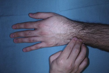
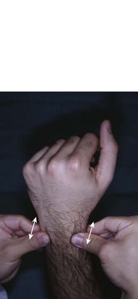
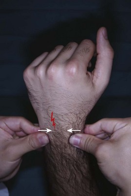
 DRU joint instability may coexist with other causes of ulnar-sided wrist pain. These include extensor carpi ulnaris (ECU) tendinitis, flexor carpi ulnaris (FCU) tendonitis, ECU subluxation, ulnar impaction syndrome, lunotriquetral (LT) instability, and pisotriquetral arthritis. It is important to consider these conditions before attributing the symptoms to DRU joint instability. The following clinical tests are suggestive of DRU joint instability:
 It is also important to rule out the presence of DRU joint arthritis because it represents a contraindication to ligament reconstruction. The following clinical test is useful in determining the presence of DRU joint arthritis.
 The patient should be examined for the presence of the palmaris longus (PL) to be used as a tendon graft.
Imaging
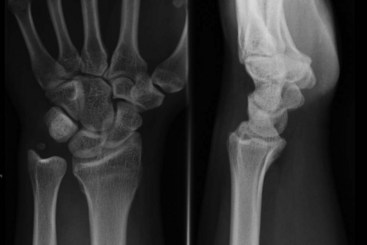
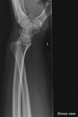
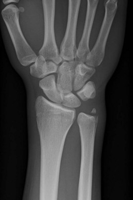
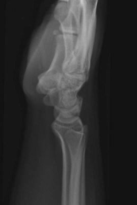
 A posteroanterior view radiograph may show widening of the DRU joint, and the lateral view demonstrates the ulnar head dorsal or volar to the radius (Fig. 82-4). A weighted lateral view (taken with patient holding 5 to 8 pounds of weight) can be used to demonstrate the instability that occurs only on loading (Fig. 82-5). Other indirect signs of instability include a basal ulnar styloid fracture (Fig. 82-6) and a displaced fleck fracture from the fovea.
 A computed tomography (CT) scan is useful in determining subtle degrees of DRU joint instability. CT scans of both wrists in identical forearm positions should be done. It is also useful in ruling out DRU joint arthritis and in assessing the comptenecy of the sigmoid notch, especially in patients with joint instability following distal radius fractures.
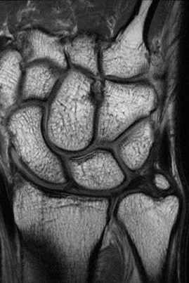
 Magnetic resonance imaging (Fig. 82-7) and arthroscopy are useful in assessment of the triangular fibrocartilage complex (TFCC) and in ruling out other causes of ulnar-sided wrist pain.
Pearls
To ensure that the lateral radiograph is adequate, the index, long, and ring metacarpals; the proximal pole of the scaphoid on the lunate; and the radial styloid in the center of the lunate should all be superimposed on one another. Additionally, the palmar surface of the pisiform should be visible midway between the palmar surfaces of the distal pole of the scaphoid and the capitate (Fig. 82-8).
Surgical Anatomy
 The DRU joint is formed by the sigmoid notch of the radius and the ulnar head. It is the distal pivot for pronosupination. This joint is stabilized predominantly by ligaments, the fibrocartilaginous lips at the rim of the sigmoid notch, and the shape of the sigmoid notch in the coronal plane.
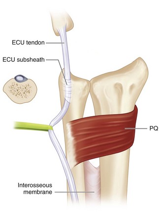
 The stabilizers of the DRU joint include the TFCC, pronator quadratus (PQ), ECU, and interosseous membrane (Fig. 82-9).
 The TFCC refers to all the soft tissues that span and support the DRU joint and ulnocarpal joints. The TFCC includes the triangular fibrocartilage (TFC, or articular disk), meniscus homologue, palmar and dorsal radioulnar ligaments, ulnar collateral ligament, ulnotriquetral ligament, ulnolunate ligament, ECU subsheath, and prestyloid recess.
 The palmar and dorsal radiolulnar ligaments are the main stabilizers of the DRU joint. These ligaments extend from the palmar and dorsal distal margins of the sigmoid notch and converge in a triangular configuration to attach to the ulna. Each radioulnar ligament divides in the coronal plane into a deep limb that inserts into the fovea and a superficial limb that inserts into the midportion of the ulnar styloid (Fig. 82-10).
Stay updated, free articles. Join our Telegram channel

Full access? Get Clinical Tree


