Fig. 1
Pre-operative anteroposterior (a) and lateral (b) view radiographs (Copyright 2014, Rubin Institute for Advanced Orthopedics, Sinai Hospital of Baltimore)
3 Preoperative Problem List
Femoral segmental bone defect with possible chronic osteomyelitis
Knee contracture secondary to rectus femoris contracture
Ankle equinus
4 Treatment Strategy
The rod was exchanged with an antibiotic cement-coated rod to prevent infection recurrence and to improve bone stability. Bone graft was mixed with vancomycin and BMP-2. Vancomycin was mixed with the bone graft because it prevents infection and does not inhibit bone healing. The bone graft was then inserted to heal the atrophic nonunion and improve bone biology. The knee was manipulated under anesthesia.
5 Basic Principles
Basic principles for the treatment of this case included improving bone stability by reaming and inserting a larger-size rod. Bone viability was also addressed by burring the bone ends at the nonunion to clean, healthy, bleeding bone. Adding bone graft obtained using the Reamer/Irrigator/Aspirator (RIA) System (DePuy Synthes, West Chester, PA) also improves the bone biology with live autogenous bone cells, and adding BMP-2 improves the recruitment of the bone healing cascade. Adding a mesh graft around the bone graft maintains the graft at the area of nonunion but at the same time allows blood and live tissue to interact with the graft since the mesh is absorbable and has large pores.
6 Images During Treatment
See Figs. 2, 3, and 4.


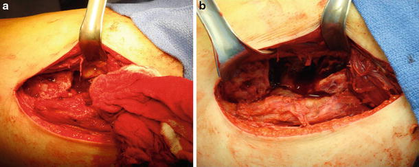
Fig. 2
Intra-operative photographs. (a) Atrophic nonunion site. (b) Resection to bleeding bone (Copyright 2014, Rubin Institute for Advanced Orthopedics, Sinai Hospital of Baltimore)
Stay updated, free articles. Join our Telegram channel

Full access? Get Clinical Tree


