Procedure 79 External Fixation of Comminuted Intra-articular Distal Radius Fractures
Indications
 Unstable unreducible extra-articular distal radius fractures
 Displaced intra-articular distal radius fractures that can be reduced by closed or percutaneous means
 Highly comminuted distal radius fractures that are reduced openly and fixed with plates or pins that require unloading of the carpus to allow healing
Examination/Imaging
Imaging
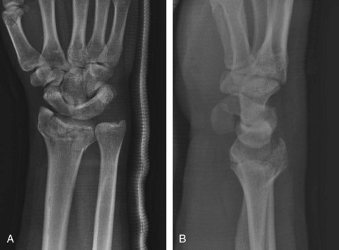
 Anteroposterior and lateral images of the wrist should be taken; these will demonstrate the nature of the fracture (Fig. 79-1).
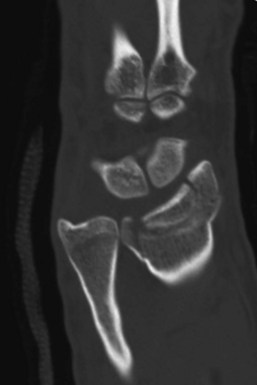
 If there is significant intra-articular comminution, a computed tomography scan will provide more information on the location and displacement of the fracture fragments (Fig. 79-2).
Surgical Anatomy
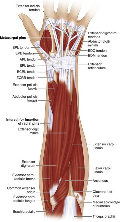
 The external fixator is placed dorsoradially across the wrist. The distal pins are placed in the second metacarpal, the proximal pins are placed in the radial shaft about a hand’s breadth proximal to the radial styloid. This position is just proximal to the “outrigger” muscles—abductor pollicis longus (APL) and extensor pollicis brevis (EPB)—as they cross the radial shaft (Fig. 79-3).
Exposures
 The first pin of the external fixator is placed in the radial shaft, and the second pin is placed in the second metacarpal. After alignment of the wrist is established, the external fixation bar slides over these two pins. The remaining pins for the metacarpal and the radius can be inserted by marking the appropriate position on the arm to permit smooth fitting of all the pin slots on the external fixator.
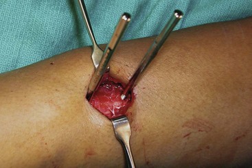
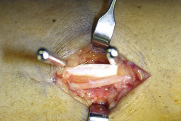
 The radial shaft pins are placed through a 3-cm incision on the “bare area” of the radius, proximal to the first compartment tendons in a dorsoradial position. The deep interval is between the extensor carpi radialis longus (ECRL) and the brachioradialis (BR) tendons. The radius should be directly visualized as the pins are placed into the bone (Fig. 79-4)
 The superficial radial nerve (SRN) needs to be visualized and protected in the proximal exposure (Fig. 79-5).
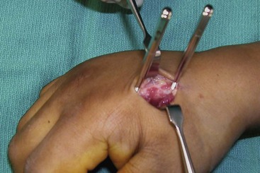
 The second metacarpal pins are placed on the dorsoradial aspect. The extensor tendons and first dorsal interosseus (FDI) muscle need to be retracted and protected (Fig. 79-6).
Procedure
Step 1
 The pins for the external fixator are placed in a dorsoradial position, about 45 degrees from radial midlateral. This position prevents the fixator from obstructing postoperative radiographs and allows the thumb full range of motion (ROM), particularly extension (Fig. 79-7).
Stay updated, free articles. Join our Telegram channel

Full access? Get Clinical Tree


