Procedure 78 Dorsal Plate Fixation and Dorsal Distraction (Bridge) Plating for Distal Radius Fractures
Dorsal Plating of Distal Radius Fractures
Examination/Imaging
Clinical Examination
 Initial examination is usually performed in the emergency department and needs to include mechanism and time of injury, hand dominance, medical comorbidities that may alter treatment, and occupation.
 Assess whether the fracture is an open or closed injury.
 Thorough musculoskeletal examination, especially the ipsilateral extremity, is necessary to rule out concomitant injuries.
 Identify any associated neurovascular and soft tissue injuries.
 Identify whether patient has any sign or symptoms of acute median or ulnar nerve injury.
Imaging
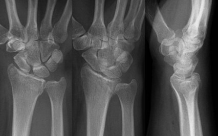
 Initial radiographic evaluation must include posteroanterior, lateral, and oblique views (Fig. 78-1).
 Assessment must include fracture angulation, displacement, radial shortening, comminution, intra-articular extension, and any ulnar-sided injuries.
 Combined lesion of a radial styloid with a dorsal marginal or facet fracture (AO type B2.2) is common and can be misdiagnosed as a Colles fracture on lateral radiographs.
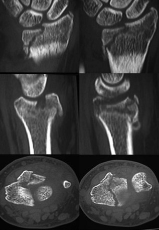
 The lunate facet often has a coronal fracture line that separates this facet into dorsal and volar fragments.
 Computed tomography scan provides excellent delineation of intra-articular extension as well as characterization of fracture comminution for preoperative planning (Fig. 78-2).
 There is also typically subchondral collapse of the lunate facet that must be addressed to ensure articular reduction.
 Proximal injuries must be ruled out by physical examination and radiographs of the elbow.
Surgical Anatomy
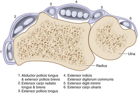
 Knowledge of the anatomic relationships of the extensor retinaculum, six dorsal extensor compartments, and convex dorsoradial cortex is essential for understanding surgical approaches as well as placement of implants on the dorsum of the radius (Fig. 78-3).
 Extensor retinaculum prevents the extensor tendons from dorsal displacement (bowstringing) and divides the tendons into six extensor compartments by vertical septae.
 The extensor pollicis longus (EPL) tendon, which lies in the third dorsal compartment, passes ulnar to the Lister tubercle and is mobilized during exposure of the dorsal distal radius.
 The extensor indicis proprius tendon and the extensor digitorum communis tendon lie in the fourth dorsal compartment and are elevated subperiosteally to minimize tendon contact with dorsally placed implants.
 Elevation of the second dorsal compartment, which contains the radial wrist extensors, puts the dorsal sensory branch of the radial nerve and the dorsal radial artery at risk, particularly if the dissection is extended distally.
 The terminal branch of the posterior interosseous nerve lies on the floor of the fourth dorsal compartment along its radial side; it can be sacrificed, if necessary, without clinical consequence.
 The articular surface of the distal radius is biconcave and triangular, with the surface divided into two hyaline covered, concave facets for articulation with the scaphoid and lunate.
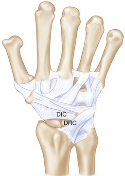
 There are two dorsal ligaments that are intimately associated with the dorsal capsule: the dorsal radiocarpal (DRC) (radiotriquetral) and dorsal intercarpal (DIC) (scaphotriquetral) ligaments (Fig. 78-4).
Exposures
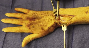
 The dorsal distal radius is approached through a straight, longitudinal incision in line with the third metacarpal and centered just ulnar to the Lister tubercle, between the third and fourth dorsal compartments (Fig. 78-5).
 Care is taken to identify and avoid injury to the superficial branch of the radial nerve.
 Full-thickness flaps containing the skin, subcutaneous tissues, and superficial fascia are raised together to expose the extensor retinaculum and the third and fourth dorsal compartments.
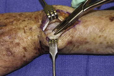
 The third dorsal compartment is longitudinally incised to mobilize the EPL tendon (Fig. 78-6).
 After mobilizing the EPL, the retinaculum is divided between the septa of the third and fourth dorsal compartments.
 Using a periosteal elevator or sharp dissection, the fourth compartment is then elevated subperiosteally from the dorsum of the distal radius, keeping the deep surface of the compartment intact.
 The distal radius is exposed sharply by elevation of the periosteum.
 Exposure of the intermediate column requires dissection to continue in an ulnar direction to the distal radioulnar joint (DRUJ).
 A longitudinal capsulotomy may be needed to expose the distal radius articular surface, which requires detaching the dorsal capsular insertions on the distal nonarticular lunate.
Pearls
Large, longitudinal veins should be preserved if possible, but crossing veins may be divided.
Full-thickness flaps will contain the dorsal sensory branches of the ulnar and radial nerve and protect them.
Subperiosteal elevation of the fourth compartment minimizes implant contact with the extensor tendons.
Pitfalls
If the distal extension of the incision is past the base of the third metacarpal, the dorsal sensory branches of both the radial and ulnar nerves are at risk.
The EPL tendon is left above the retinaculum at closure to minimize the risk for tendon injury by ischemia or direct contact with an implant.
Care must be exercised not to enter the DRUJ. If the dorsal radioulnar ligament is divided during ulnar dissection, radioulnar instability can result.
The intercarpal ligaments must be protected during capsulotomy.
Procedure
Dorsal Plate Fixation for Distal Radius Fractures
Step 1: Reduction of Dorsally Angulated Fractures
 Once the proximal fracture lines and articular surface are exposed and assessed, all hematoma is evacuated.
 Lister tubercle is removed with a rongeur or small osteotome (Fig. 78-7).
 Reduction of the dorsal angulation can be performed with the use of narrow-angled Homan retractors along the proximal fragment.
 With gentle traction and lifting of the bone retractors, reduction of the dorsal angulation can be achieved.
 Provisional fixation with Kirschner wires (0.045-in.) can be used to hold the reduction and is checked fluoroscopically.
Step 2: Fixation of Dorsally Angulated Fractures
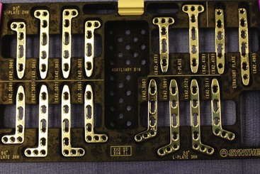
 After satisfactory reduction is performed, a 2.4-mm low-profile dorsal T-plate (Fig. 78-8) can be placed directly over the dorsal surface of the distal radius.
 Preliminary fixation of the plate with a single, bicortical compression screw placed in the oblong hole of the plate will allow for proximal-distal adjustments in plate position as determined clinically and fluoroscopically.
 It is important to provisionally bend the plate to fit the contour of the dorsal radial cortex so that the plate lies flush with the dorsal cortex.
 Although several options for plating are available, including locking and nonlocking plates, one must consider contouring a fixed-angle plate by threading a locking guide into the plate before contouring; this will avoid damage to the threads of the locking mechanism.
 The final placement is checked by fluoroscopy in anteroposterior and lateral views to confirm proper positioning of the plate.
 Three to five 2.7-mm screws are then placed bicortically along the distal row of the plate, using fluoroscopy to confirm placement.
 Two additional proximal screws are then inserted bicortically, and final fluoroscopic assessment is performed.
Step 3: Reduction and Fixation of Dorsal Marginal Fractures
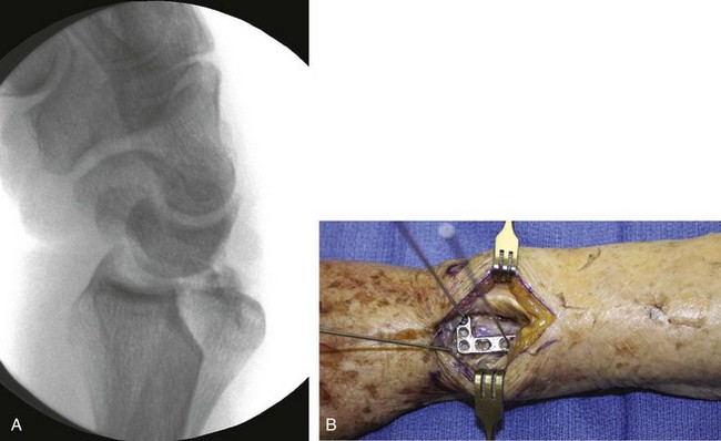
 It is important to assess carpal subluxation and initially restore the radial styloid fragment if present (Fig. 78-9A).
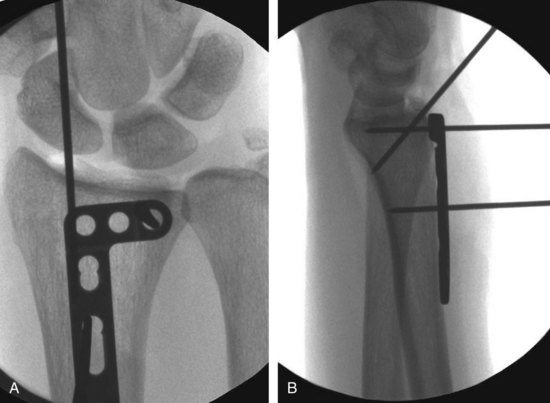
 This reduction is usually accomplished with traction and ulnar deviation of the hand and wrist, and then provisional K-wire fixation (Figs. 78-9B and 78-10).
 Once the appropriate-sized 2.4-mm plate has been contoured, the locking screw guide is placed in the proximal screw hole and is used to hold and contour the plate.
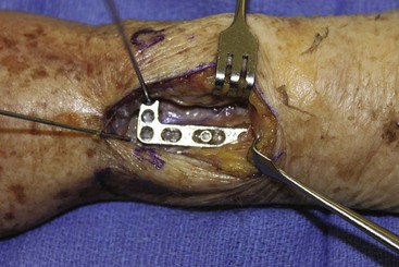
 Preliminary fixation of the plate with a single, bicortical screw placed in the oblong hole will allow for proximal-distal plate adjustments as determined clinically and fluoroscopically (Fig. 78-11).
 A provisional K-wire is then placed through the threaded locking guide at the distal end of the plate but not into the radius to facilitate quick placement after reduction of the carpus.
 The dorsal lip or marginal fracture can be reduced now against the scaphoid and lunate, which will correct any associated dorsal carpal subluxation.
 The marginal fracture will then be provisionally fixed into the volar cortex through the provisional K-wire that had been previously placed into the threaded locking guide at the distal end of the plate.
 Congruity of the articular surface must be confirmed with direct visualization as well as fluoroscopically.
 If the plate is acting purely as a buttress, distal locking screws are not necessary.
 If additional fixation for stability is necessary, 2.4-mm screws are then placed bicortically along the distal row of the plate, using fluoroscopy to confirm placement.
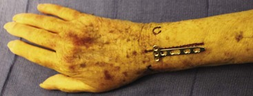
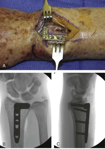
 One or two additional bicortical screws are placed proximally to the proximal extent of the fracture to finalize construct (Figure 78-12).


