Procedure 75 Scaphotrapeziotrapezoid Arthrodesis and Lunate Excision with Replacement by Palmaris Longus Tendon
Indications
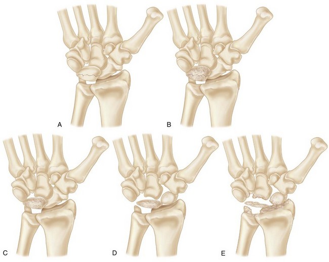
 Advanced avascular necrosis of the lunate from late stage IIIA to early stage IV according to the Lichtman preoperative 5-stage classification.
 This procedure is mainly indicated for stage IIIB Kienböck disease, which is associated with carpal collapse with a fixed palmar scaphoid flexion.
 A radiograph showing generalized arthrosis is not suitable for scaphotrapeziotrapezoid (STT) arthrodesis and lunate excision with replacement by the palmaris longus tendon.
 This procedure is contraindicated in patients with preoperative advanced osteoarthrosis between the radius and scaphoid because of the risk for further progression of osteoarthrosis at the radioscaphoid joint.
Examination/Imaging
Clinical Examination
 A complete examination of the involved and contralateral extremity is indicated, especially if the patient has a history of autoimmune disease or infection.
 Documentation of baseline pinch and grip strength is helpful.
 Attention to soft tissue swelling must be given because some patients have associated capsular inflammation, thickening, and synovitis.
 Pain can be elicited in all directions of motion, and frequently there is a notable decrease in the arc of motion, particularly in extension.
 Degree of pain, range of motion of the wrist, and grip strength of both wrists should be meticulously measured preoperatively to evaluate postoperative results.
Imaging
Plain Radiographs
 Staging: Lichtman modification of Stahl staging
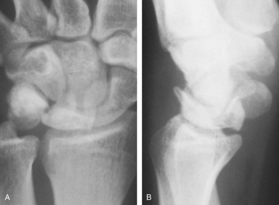
 Posteroanterior (PA) and lateral radiographs with the wrist in neutral rotation and neutral flexion-extension (Fig. 75-2).
 Ulnar variance radiographic views with the wrist in full pronation and full supination. The carpal height ratio is measured from PA view with the wrist in neutral position. The Stahl index is also measured from lateral view.
 Lunate sclerosis, carpal instability patterns, and fragmentation are seen.
 Shortened scaphoid in the long axis and cortical ring sign of the scaphoid are seen in stage IIIB, which indicates scaphoid in flexed position.
 Evaluation of relationship among scaphoid, trapezium, and trapezoid.
Computed Tomography
 Computed tomography (CT) is needed to evaluate precisely the bony architecture of the lunate and the remainder of the carpus to determine which procedure is indicated and to determine whether arthritic changes of the midcarpal and radiocarpal joints are present.
 A CT scan is useful for evaluation of fragmentation or fracture patterns and associated collapse.
Magnetic Resonance Imaging
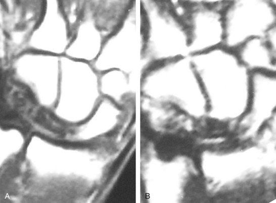
 MRI will show decreased uptake in both T1- and T2-weighted images. Gadolinium contrast has improved diagnosis and evaluation of lunate vascularity (Fig. 75-3).
 It is important to differentiate between ulnar impaction of the lunate and true Kienböck disease.
 Stage 0 has been proposed when lunate edema is visible on MRI only. MRI can show the degree of devascularization or revascularization.
Stay updated, free articles. Join our Telegram channel

Full access? Get Clinical Tree


