Procedure 72 Dorsal Capsulodesis for Scapholunate Instability Using Suture Anchors
![]() See Video 53: Dorsal Capsulodesis for Scapholunate Ligament Injury
See Video 53: Dorsal Capsulodesis for Scapholunate Ligament Injury
Indications
 Static or dynamic dorsal intercalated segmental instability (DISI) deformity is found without a wide scapholunate (SL) gap.
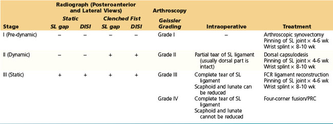
 We determine the treatment for SL instability based on clinical presentation, radiographic appearance, and arthroscopic and intraoperative findings. Our current algorithm for selection of appropriate procedure for SL instability is indicated in Table 72-1.
Examination/Imaging
Clinical Examination
 Patients complain of radial-sided wrist pain especially with loading activities, weakness of grip, and swelling, along with discomfort at extremes of wrist extension and radial deviation.
 Tenderness is found in the radial snuffbox or over the SL interval just distal to the Lister tubercle.
 The following two provocative tests have been described for global assessment of wrist pain. These tests are useful in patients when the history and findings of examination do not match. However, they do not indicate the location or nature of the pathology.
 One or more of the following provocative tests may be positive depending on the degree of injury to the SL ligament and integrity of the secondary stabilizers.
Imaging
 Stress views, including the clenched-fist view, posteroanterior maximal radial deviation, and posteroanterior maximal ulnar deviation, are useful in the diagnosis of dynamic SL instability.
 Arthroscopic examination has become the standard method of evaluation of interosseous ligamentous injuries. The arthroscopic classification of ligament injuries proposed by Geissler (Table 72-2) is useful in determining treatment options.
Surgical Anatomy
 The SL ligament is a C-shaped structure that connects the dorsal, proximal, and volar surfaces of the scaphoid and lunate. Partial SL ligament tears most often involve only the proximal and volar components of the SL interosseous ligament complex. The dorsal ligament is much stronger than its volar counterpart and may resist substantial traumatic torques without yielding.
 The normal kinematics of the SL joint is governed by the SL ligament, a primary stabilizer, and by an envelope of surrounding extrinsic ligaments that act as secondary stabilizers, namely the radioscaphocapitate (RSC), the long and short radiolunate (LRL and SRL) ligaments on the volar side, and the dorsal radiocarpal (DRC) and dorsal intercarpal (DIC) ligaments on the dorsal side.
 When the scaphoid has lost its connections to the lunate, the role of secondary stabilizers is particularly important. Their failure results in the static carpal malalignment.


