7: Tissue Assessment, Decision Processes, and Operative Planning

Chapter 7 Tissue Assessment, Decision Processes, and Operative Planning


Background and Importance of Tissue Based, Quantitative Systems and Decision Processes


Breast augmentation outcomes are largely determined by preoperative decisions. The quality of preoperative decisions is directly related to the information on which the decisions are based and the prioritized decision making processes that surgeon and patient use preoperatively. This chapter focuses on the logic and published data that validate tissue based, quantitative preoperative patient assessment and decision processes, and the clinical application of those processes.


Preoperative Parameters and the Scientific Validity of Breast Augmentation Studies


Scientific validity of any prospective or retrospective study in breast augmentation requires that a study address variables that affect outcomes, defining those variables preoperatively, collecting objective, quantified data, and analyzing that data to test a hypothesis. A consistent weakness of most published studies in breast augmentation is a failure to define critical variables preoperatively and collect objective data pre- and postoperatively that relate to those variables. More than 53 variables affect the outcome of every breast augmentation. Table 7-1 lists surgeon and implant variables that exist in every breast augmentation.1


Table 7-1 Variables that impact outcomes in breast augmentation


Clinical and tissue variables Surgeon variables
Genetic factors Incision approach
Hormonal factors
Pregnancy history
Nursing history Pocket dissection method
Age Pocket dissection technique
Medications (especially birth control pills and hormones) Degree of tissue trauma
Skin and subcutaneous tissue thickness
Skin compliance


Postoperative drug regimen
Size of periprosthetic pocket
Shape of periprosthetic pocket
Patient arm position


Implant fill volume
Air in implant
TOTAL TISSUE VARIABLES: 31 TOTAL SURGICAL VARIABLES: 22
TOTAL VARIABLES, TISSUES AND SURGICAL: 53


The extent to which surgeons address these 53 variables by recording objective, quantifiable pre- and intraoperative data for subsequent analysis largely determines the scientific validity of any clinical breast augmentation study. If a study fails to address these variables preoperatively, no amount of sophisticated statistical postoperative analysis can create scientific validity. Any statistical analysis is only as scientifically valid as the data which the process analyzes. A majority of published studies on breast augmentation are scientifically weak because of failure to address critical preoperative variables.


To be scientifically valid, any study that compares methodologies, surgical techniques, or implants must prospectively create comparative cohorts of patients that acknowledge and control for the 53 variables listed in Table 7-1. Regardless of the methods, techniques or implants the study attempts to compare, conclusions are scientifically invalid if the investigator does not preoperatively define two groups of patients, with each of the two groups having similar variables to those listed in Table 7-1. Addressing a small percentage of these variables is not adequate to validate conclusions—the two groups must have similar age, pregnancy, and tissue characteristics, quantified objectively, to achieve scientific validity.


Impact of Surgeon Education on Patients’ Experience and Outcomes


Surgeon education in breast augmentation and many other surgical disciplines is based on a preceptor model. Resident plastic surgeons learn breast augmentation principles and techniques from their professors and attending physicians. In many academic plastic surgery programs, resident experience in breast augmentation is limited, and attending academic surgeons may perform very few breast augmentations compared to plastic surgeons in private practice. When academic and private practice surgeons who teach residents refer to breast augmentation as a “simple” operation, it is not surprising that for more than three decades, reoperation rates of 15–20% in just 3 years24 following breast augmentation have changed minimally, and for many patients, the patient experience (recovery and outcomes) remains largely unchanged.


Improvement in patient outcomes in breast augmentation relates directly to the development and dissemination of improved methodologies and scientifically proved processes. The extent to which surgeon residency education programs and professional society education programs effectively disseminate new information about proved processes and the extent to which surgeons implement proved processes determine future improvements in patient outcomes. Improving patient outcomes requires that those who define content for educational programs or venues prioritize dissemination of proved processes that have objectively documented superior outcomes in peer reviewed and published studies. When surgeons use attendee evaluation form data and prioritize maximizing numbers of surgeons on a program with short presentations in lieu of thorough delivery of proved process content, the result is often more entertaining than educational. Ultimately, the degree to which surgeons choose to learn, improve, and implement proved processes, allocating time and financial resources, determines the experience and outcomes of their patients.


Redefining the Patient Experience in Breast Augmentation


The current decade has produced quantum improvements in surgeons’ approach to breast augmentation and has redefined the potential patient experience, outcome, and reoperation rates. Advances in tissue assessment, preoperative decision processes, surgical techniques, implant devices, and postoperative management now allow surgeons to routinely enable patients to return to full normal activities within 24 hours following breast augmentation with an overall reoperation rate of 3% over 7 years postoperatively.57 For the first time in the history of Food and Drug Administration (FDA) premarket approval (PMA) studies of breast implants, refined decision processes, methodologies, and techniques produced a zero percent reoperation rate in 50 consecutive patients in an FDA study of Inamed/Allergan Style 410 form stable, cohesive gel anatomic breast implants.8 These methods, which have redefined the surgeon and patient experience in breast augmentation, are peer reviewed and published in the journal Plastic and Reconstructive Surgery, and are available to surgeons worldwide. The specific tissue assessment, preoperative decision processes, and operative planning methodologies are the subject of this chapter.


Evolution of Preoperative Planning and Implant Selection Methods


In plastic surgery, artistry is an invaluable adjunct to science. Artistry is a complement to science, not a substitute for quantifiable objectivity and the scientific method. Even Michelangelo and Leonardo—consummate artists—were consummate believers in the value and necessity of extensive measurements and planning before embarking on any artistic endeavor.


For decades, surgeons and patients have based choices and decisions about breast implant size and type on subjective parameters—patients’ wishes for a particular bra cup size, requests to look like a particular photograph from a magazine or another patient’s photographic outcome, or a volume to mimic a test implant placed into a bra. While each of these methods may appeal to some patients and surgeons, none is quantifiable, none is objective, and none is scientific.


Optimizing outcomes for patients requires developing processes of decision making that can be refined, optimized, and transferred to other surgeons—systems that use quantifiable parameters to make objective decisions, providing data that can be used to scientifically test hypotheses. A measurement system for breasts is similar to other measurement systems in common use in plastic surgery—systems that define optimal dimensions for cleft lip or palate repair, craniofacial surgery, and virtually every reconstructive procedure where quantifying a dimension helps a surgeon deliver a more accurate product.


While plastic surgeons routinely use and insist on quantifiable parameters in a wide range of reconstructive and cosmetic procedures, a relatively small percentage of surgeons currently base decisions in breast augmentation on quantifiable tissue dimensions and parameters. Quantitatively based systems do not dictate to surgeons or patients what they can choose, nor do they dictate a standard of practice. Systems for implants based on quantifiable parameters to define individual patient tissue characteristics offer surgeons additional tools to help make decisions more objective than subjective, make outcomes more predictable, and reduce reoperation rates.


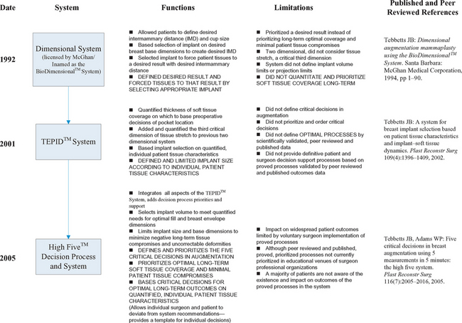
Table 7-2 summarizes the evolution of quantitative, tissue based systems and decision process support for patient assessment and critical decisions in breast augmentation since their inception in 1992.


Table 7-2 Evolution of quantitative, tissue based systems and decision process support for patient assessment and critical decisions in breast augmentation.


Objectives and Limitations of the First Generation Dimensional System


To attempt to increase the reliability and predictability of results to patients, in 1990 the author developed a dimensional system that enabled a more quantitative method of assessing a patient’s breasts and selecting appropriate implants to deliver the patient’s desired result. This first dimensional system was published in a monograph in 1994,9 and was licensed by McGhan Medical Corporation as the BioDimensional™ System and subsequently distributed worldwide. During the past decade, the BioDimensional™ System has become the most widely accepted and used dimensional system worldwide by providing surgeons with a more objective, quantitative, and scientific approach to breast evaluation and decision making in breast augmentation.


When first introduced in 1994, the BioDimensional™ System9 allowed patients to define their desired intermammary distance by displacing the breasts medially, then using measurements of the existing breast width and the desired breast width to determine the base width of the implant required to deliver the patient’s desired result. The original dimensional system functioned in two dimensions (base width and height), and focused on forcing tissues to a desired result (delivering the desired base width to achieve the patient’s goals for desired intermammary distance). This system is still in use as the Allergan BioDimensional™ System. This first generation dimensional system produced a paradigm shift in surgeons’ approach to patient assessment and decision making in breast augmentation. For the first time, surgeons could design a desired result using a quantitative assessment and decision making methodology instead of relying on bra stuffing, photographs, or subjective cup size methods to define objectives.


While this first generation system reoriented many surgeons’ thinking toward a more quantitative, reproducible, scientific approach to patient assessment and implant selection, the system has evolved two more generations with more than a decade of experience in a wide range of breast types. The strengths of the first generation system were (1) attempting to quantify dimensions and patient tissue characteristics, (2) basing decisions on quantifiable parameters, (3) enabling more scientific evaluation of quantified parameters, and (4) encouraging more consistent and predictable results.


An additional decade of clinical experience and longer-term followup of larger numbers of patients have pointed out limitations of this first generation system: (1) the system prioritized achieving a specific result instead of prioritizing optimal soft tissue coverage long-term (did not include specific, quantifiable parameters that mandate optimal tissue coverage, (2) the system and current revisions by other surgeons have no guidelines or restrictions on volume limits, (3) the system is two dimensional, omitting a critical third dimension—tissue stretch—that affects decisions about optimal volume for a specific patient’s envelope and provides information about risks of excessive stretch with bottoming or traction rippling, and (4) the system was originally designed to be specific to one manufacturer’s family of anatomic implant products.


In summary, the first generation BioDimensional™ System allows patients and surgeons to design a desired result by width and height dimensions, and then select an implant of appropriate dimensions to force the tissues to the desired result. In contrast to the first generation system, next generation systems do not force tissues to a desired result; instead, they recognize and quantify what the tissues will allow or what the tissues require to achieve an optimal long-term result with minimal negative tissue consequences.


Priorities for the Next Generation Measurement and Decision Process System


Priority 1 is preserving the patient’s tissues over time by making optimal decisions of volume and coverage based on quantifiable parameters, not prioritizing an arbitrary implant size that may compromise the patient’s tissues over time. An optimal system for tissue analysis and implant selection should prioritize the quality and integrity of patients’ tissues long-term above all other considerations by defining quantifiable guidelines for total weight (volume) of the breast implant based on tissue coverage considerations. Patients often arrive at their consultation “knowing” what they want, and one of the responsibilities of the surgeon is to meet a patient’s expectations. An equally important question, however, is whether the patient is adequately educated to understand how what she wants is likely to affect what she is realistically likely to get long-term—the potential long-term tissue consequences of her current wishes. Absent guidelines built into a system that integrates patient education and informed consent systems with quantifiable tissue assessment, patients and surgeons will likely continue to select implant size and type based on subjective and arbitrary considerations of breast size without acknowledging responsibility for the potential longer-term tissue consequences of their choices. The ultimate goal for the welfare of the patient is prioritizing her tissues over her wishes if she desires an optimal long-term result with minimal tissue compromises, reoperations, and uncorrectable tissue deformities.


To avoid implant palpability, visibility, visible traction rippling, and extrusion risks long-term, the surgeon must prioritize optimal, long-term soft tissue coverage of the implant and preservation of the patient’s existing breast parenchyma long-term, above all other considerations. Some of the most preventable complications of breast augmentation occur when surgeons and patients fail to prioritize optimal soft tissue coverage of the implant or decide to select implants with excessive size or projection that stretch, thin, and compromise tissues, and increase risks of parenchymal atrophy long-term.


Priority 2 is to provide quantitative data regarding patient tissue characteristics—data that enable surgeons to make objective decisions and allow scientific evaluation of outcomes.


Priority 3 is that all measurements concerning implant weight (size, volume) and coverage be made on the breast, not from landmarks on the breast to other landmarks on the chest, torso, or abdomen. The ultimate consequences and aesthetics of any breast implant to the tissues of the breast are within the boundaries of the breast. When performing primary augmentation, surgeons do not relocate the breast on the torso. While some surgeons may be tempted to base implant dimensions (especially implant height) on measurements such as sternal notch-to-nipple distance, these measurements and the subsequent decisions may be overly simplistic and inaccurate, depending on the filler distribution dynamics and implant–soft tissue dynamics of the implant selected.


Principles of Breast Aesthetics and Implant–Soft Tissue Relationships


Specific characteristics and relationships define an aesthetic breast. Every breast has a specific amount of skin that defines the dimensions—base width, stretch, and height—of the skin envelope. The skin envelope has specific tissue characteristics—base width, thickness, and stretch—that determine how much the envelope will stretch in response to adding fill (an implant). A funnel analogy is helpful when discussing these issues with patients. If the surgeon could pour filler into a funnel in the top of the breast, the lower breast would fill first until it reached its limit of anterior stretch, then the middle portion of the breast would fill, and finally the upper pole of the breast would fill to create an optimal breast contour and optimal transition of the breast from the chest. If the surgeon adds inadequate fill to any breast, the lower breast will be fuller compared to the middle or upper breast. When the breast is full with an optimal contour, adding additional volume creates excessive fullness in the upper breast, desired by some patients and surgeons, but also adding weight that ultimately causes more stretching of the lower envelope and loss of upper fill.


The wider the base width (BW) of the existing parenchyma, and the greater the skin stretch measured by anterior pull skin stretch (APSS), the greater volume required to fill the envelope for optimal aesthetics. Two components define optimal fill postoperatively: the patient’s existing parenchyma and the implant. To predict optimal implant volume, the surgeon must estimate the parenchyma’s contribution to stretched envelope fill (PCSEF). If the envelope is adequately filled preoperatively with the patient’s existing parenchyma (>80%) and the skin stretches minimally, less volume is required in the implant. Conversely, if the skin stretches significantly, and minimal parenchyma (<20%) is present, more implant volume is required for optimal fill of the envelope.


Optimal fill is defined as the minimal amount of fill required to produce an optimal aesthetic result while minimizing potential negative tissue consequences. In a parous breast, more volume is often required due to the width or increased stretch characteristics of the envelope. Conversely, in a nulliparous breast with APSS < 2 cm (a tight envelope) and PCSEF > 80% (an envelope already full with parenchyma), much less volume is required for optimal fill.


When selecting an implant device, surgeons must consider not only the effects of volume/weight, but also the effects of focusing that volume in a specific area to achieve projection. For any given base width breast implant, increasing projection means adding weight and adding pressure directly behind the existing parenchyma (usually to satisfy a patient or surgeon’s desire for a more projecting breast). Pressure focused on lower pole parenchyma can produce parenchymal atrophy over time. Currently there are no scientifically valid studies that define amounts of parenchymal atrophy that occur with varying projection implants, but experienced surgeons who have long-term followup of more than 10 years with highly projecting or high profile devices are aware that highly projecting implant devices can have negative tissue consequences to the parenchyma and lower pole skin envelope over time that include irreversible skin stretch and parenchymal atrophy. A high profile implant may temporarily create a more projecting breast, but lose the projection over time due to parenchymal atrophy, resulting in a permanent, uncorrectable compromise to the patient’s tissues with loss of parenchyma.


The wider the base width of a patient’s parenchyma, the longer the nipple-to-inframammary fold measurement for optimal aesthetics. Stated another way, the optimal position of the inframammary fold depends on the width of the breast. If the breast is excessively wide compared to the nipple-to-fold measurement, the breast appears boxy. If nipple-to-fold distance is too great compared to the width of the breast, the breast appears narrow, tubular, or ptotic. When increasing the width of the breast during augmentation, the optimal level to set the inframammary fold intraoperatively depends on the planned base width of the postoperative breast.


Two Basic Approaches to Breast Augmentation


Surgeons currently use two basic approaches to breast augmentation: (1) defining a desired result (using dimensions or subjective parameters) and selecting implants to force the tissues toward the desired result, and (2) allowing the tissues to define optimal fill by the dimensions and stretch characteristics of the envelope.


The first approach—forcing the tissues to a desired result—often produces larger breasts, especially in younger, nulliparous patients, compared to the latter, by forcing tighter tissues to stretch more than they stretch under manual measurement (APSS), and by widening the breast using an implant with a base width that exceeds the base width of the existing parenchyma. When patients and surgeons choose this approach to achieve a desired result, patients should be aware and acknowledge in informed consent documents that forcing their tissues to the desired result may produce negative tissue consequences long-term, including tissue stretch and thinning, parenchymal atrophy, visible or palpable implant edges, visible traction rippling, and ptosis. Some of those tissue consequences may be irreversible.


The second approach—allowing the tissues to define optimal fill by the dimensions and stretch characteristics of the envelope—attempts to minimize negative tissue consequences long-term in a wide variety of breasts. The simplest of approaches—selecting an implant with a base width less than the base width of the existing parenchyma and a projection less than or equal to the limit of manual stretch of the breast skin (APSS)—can avoid a very large number of uncorrectable tissue consequences and complications of breast augmentation.


No system is without tradeoffs. Even with a system that allows tissue dimensions and stretch to define volume requirements, some wide, parous breasts with minimal to moderate parenchyma require very large implants in order to adequately fill the envelope for an optimal aesthetic result. In these cases, patients must be aware that the amount of volume required for an optimal result may add enough weight to cause negative consequences to their tissues over time such as tissue stretch and thinning, parenchymal atrophy, visible or palpable implant edges, visible traction rippling, and ptosis. Some of those tissue consequences may be irreversible.


Tissue Coverage Priorities and Measurements


Assuring optimal soft tissue coverage requires surgeons to quantify soft tissue coverage in critical areas over the implant and use quantified criteria for pocket selection and additional soft tissue cover.


Traditional methods of subjective tissue assessment (e.g. thick, thin, tight, loose) are impossible to quantify, and therefore cannot be used to make objective decisions based on criteria determined by data. Without quantified criteria, surgeons cannot scientifically evaluate outcomes and derive outcomes based treatment algorithms.


Specific measurements of tissue coverage parameters should include measurements of pinch thickness of skin and subcutaneous tissue superior to the breast parenchyma (STPTUP, soft tissue pinch thickness of the upper pole) and at the inframammary fold (STPTIMF). Patients whose tissues are thin (less than 2 cm pinch thickness above the existing parenchyma) are at risk short- and long-term for visible edges of the implant superiorly and superior traction rippling caused by traction of the implant on the capsule attached to the thin overlying soft tissues.


The system should set a quantified criterion to suggest additional muscle coverage (e.g. STPTUP < 2 cm) by placing the implant in either a traditional partial retropectoral or a dual plane (dividing origins of pectoralis along the inframammary fold) position. The system should also set a quantified criterion that suggests preserving muscle origins of the pectoralis intact along the inframammary fold when patient tissues along the fold are exceedingly thin (e.g. STPTIMF < 0.5 cm), choosing a traditional subpectoral pocket instead of a dual plane pocket that divides pectoralis origins along the inframammary fold.


To assure optimal pectoralis coverage medially when additional coverage is indicated, the system should remind surgeons to avoid any division of pectoralis origins medially along the sternum, from the xiphoid inferiorly to the junction with the medial inframammary fold.


Most importantly, the system should always specify an implant base width that is less than or equal to the base width of the patient’s existing parenchyma in order to assure parenchymal coverage of implant edges. If surgeons applied only a single dimensional criterion when selecting an implant, this criterion alone could avoid many complications that occur as the result of inadequate tissue coverage. The only exception to this specification is in very narrow base width breasts with base widths less than 11 cm, including constricted lower pole breasts and tubular breasts. In these cases, widening the base width of the breast may be required for optimal correction of the deformity, but the patient should understand and accept the potential coverage compromises that might occur in the future and acknowledge these possible consequences in informed consent documents preoperatively.

< div class='tao-gold-member'>

Stay updated, free articles. Join our Telegram channel

Jul 23, 2016 | Posted by in Aesthetic plastic surgery | Comments Off on 7: Tissue Assessment, Decision Processes, and Operative Planning

Full access? Get Clinical Tree

Get Clinical Tree app for offline access