Procedure 69 Vascularized Bone Grafting for Scaphoid Nonunion
![]() See Video 51: Pedicled Vascularized Bone Transfer for Scaphoid Nonunion
See Video 51: Pedicled Vascularized Bone Transfer for Scaphoid Nonunion
Indications
 Scaphoid fracture nonunions are seen that have developed avascular necrosis (AVN).
 Vascularized bone grafting is contraindicated in fracture nonunions associated with significant degenerative changes in the radioscaphoid or midcarpal joint. These wrists require a salvage procedure.
 A scaphoid nonunion humpback deformity cannot be corrected unless a structural graft is used.
Examination/Imaging
Clinical Examination
 General dorsoradial edema and focal snuffbox tenderness are highly suggestive of a scaphoid fracture.
 The distal pole of the scaphoid may also be tender to palpation, and axial compression of the thumb may reproduce the pain.
 Decreased wrist motion and grip strength are common findings in patients with a scaphoid nonunion.
Imaging
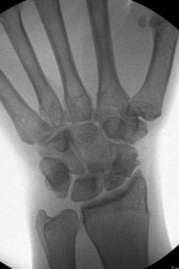
 Wrist radiographs including scaphoid views should be obtained to evaluate the overall fracture pattern, carpal alignment, and degree of radiocarpal and midcarpal arthritis. Chronic scaphoid nonunions display a characteristic pattern of wrist arthritis and carpal instability—scaphoid nonunion advanced collapse (SNAC), as described by Watson and Ballet. The hallmark of AVN on radiographs is increased bone density, fragmentation, and collapse (Fig. 69-1).
 A thin-cut computed tomography (CT) scan with reconstructions carefully aligned in the axis of the scaphoid is helpful to elucidate scaphoid angular deformity (as measured by intrascaphoid angles), translation, comminution, and degenerative changes. This study best quantifies the deformity and necessary reconstruction.
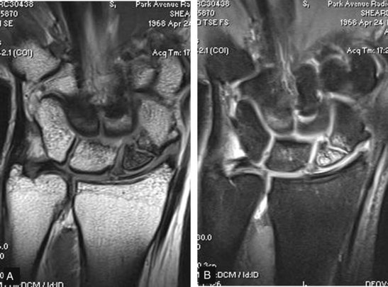
 Magnetic resonance imaging (MRI) is the only noninvasive study able to assess vascularity of the scaphoid and identify AVN (Fig. 69-2). An MRI should be obtained in every patient for whom there is a concern about the viability of the proximal pole. Contrast-enhanced imaging has a sensitivity of 66%, a specificity of 88%, and an accuracy of 83%.
Surgical Anatomy
 Eighty percent of the scaphoid surface is covered by articular cartilage, leaving limited space for vascular supply.
 A dorsal branch of the radial artery provides 70% to 80% of the blood supply.
 A second nutrient artery enters volarly and supplies only the distal pole.
 There is no nutrient artery to the proximal pole; thus, it is susceptible to AVN when fractured.
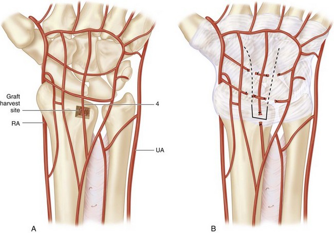
 Dorsal distal-radius vascularized bone grafts have been shown to be based on consistent vascular anatomy (Fig. 69-3A).
 The pedicled graft described in this chapter is based on the artery of the fourth extensor compartment. The fourth extensor compartment artery is less than 1 mm in diameter and between 1 and 2 cm in length (see Fig. 69-3A and B).
 Rotation of the graft on the capsular pedicle of only 10 to 30 degrees allows placement of the graft on the proximal scaphoid.


