Fig. 1
Pre-operative clinical photographs of the (a) front, (b) side, and (c) back view of the patient showing an equinus deformity of 50°, hindfoot varus of 35°, midfoot cavus, forefoot adduction and supination, as well as claw toes. Atrophy of the calf and peroneal muscle compartment characteristic of CMT is noted on the right leg. The left foot demonstrates a minor degree of cavovarus deformity
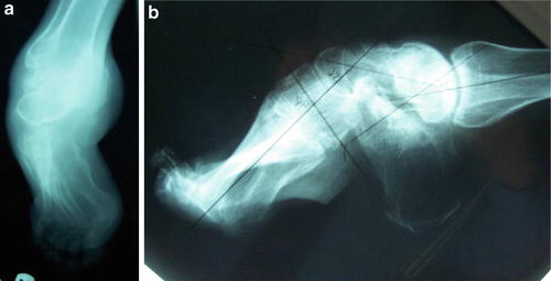
Fig. 2
Pre-operative standing ankle (a) anteroposterior (AP) and (b) lateral radiographs of the right ankle and foot demonstrating the aforementioned equino-cavovarus deformity
3 Preoperative Problem List
1.
CMT
2.
R ankle equinus with tight tendo-Achilles
3.
Hindfoot varus
4.
Midfoot cavus with tight plantar fascia
5.
Forefoot adduction and supination
6.
Claw toes
4 Treatment Strategy
1.
Release of tight tendo-Achilles contributing to the equinus contracture.
2.
Release of the plantar fascia partially contributing to the cavus deformity.
3.
Release the posterior tibial tendon, FDL, and FHL.
4.
Plan for gradual correction of both hindfoot and midfoot. However, if acute correction of the ankle/hindfoot can be done, the frame will be more simple requiring gradual correction of only the midfoot.
5.
Correction of the hindfoot varus, midfoot cavus, and forefoot adduction/supination by performing a midfoot osteotomy at the naviculo-cuboid level with gradual correction via application of TSF after the aforementioned soft tissue releases to achieve complete correction.
6.
Percutaneous flexor tenotomies to correct claw toe deformities.
5 Basic Principles
1.
Perform a tendo-Achilles lengthening for the equinus contracture through a minimal posterior incision.
2.
Through a medial approach, perform fractional lengthening of the PTT-FDL-FHL to correct hindfoot varus (if required in a non-flexible deformity as evidenced on clinical examination). In this case, after these soft tissue releases, the ankle equinus and the hindfoot varus were acutely corrected without difficulty. The TSF was used to hold the ankle and hindfoot correction and to gradually correct the midfoot.
3.
Through a plantar approach, release the plantar fascia to partially correct the cavus associated with the tight plantar soft tissue structures before proceeding with the bony element of the cavus deformity.
4.
Additional percutaneous tenotomies of the flexor tendons were performed with percutaneous K-wire fixation of the distal and proximal interphalangeal joints of the toes and transfixing the K-wires to the TSF.
5.
Application of TSF with a ring on the distal tibia and a U-ring butt frame around the hindfoot. A third ring is mounted over the midfoot distal to the anticipated osteotomy site at the naviculo-cuboid level.
6 Images During Treatment
Related posts:
 8: Femoral Bone Defect
 11: Bone Transport Over a Nail for Infected Tibial Nonunion and Bone Defect
 22: Bone Transport to a Knee Fusion and Secondary Intramedullary Nailing s/p Gunshot Wound
 78: Two-Stage Treatment of a Chronic Foot Dislocation
 84: Lapidus Fusion with External Fixation
 65: Closed Correction of Club Foot with Ilizarov
Stay updated, free articles. Join our Telegram channel

Full access? Get Clinical Tree


