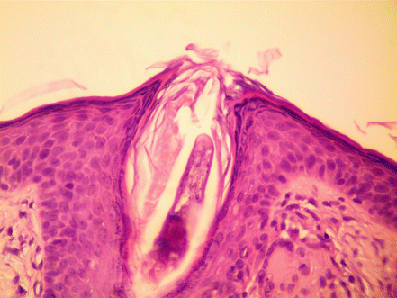
Figure 31.1
Demodex mite. A circular lesion marked by ink dots was biopsied
Clinical Differential Diagnosis
Fibrous papule
Basal cell carcinoma
Sebaceous hyperplasia
Cyst
Demodex folliculitis
Histopathology
Sections showed a portion of an insect within the follicular infundibulum. A mild perifollicular of lymphocytes and histiocytes were found (Fig. 31.2).


