Procedure 65 Arthroscopic Treatment for Septic Arthritis
Indications
 Arthroscopic synovectomy has been shown to be beneficial in patients with symptomatic, refractory reactive arthritis.
 Arthroscopy allows for not only continuous irrigation of joint space but also débridement of infected material.
Examination/Imaging
Clinical Examination
 Examine the entire wrist for swelling, redness, warmth, and pain.
 The range-of-motion examination will produce exquisite pain with both active and passive motion.
 Hematogenous spread will manifest with systemic signs of infection, which may include fever, chills, tachycardia, and diaphoresis.
 Evaluation with laboratory tests should be done and should include white blood cell count (WBC), erythrocyte sedimentation rate, and C-reactive protein levels.
 Joint fluid can be obtained at the initiation of the procedure and should be evaluated with Gram staining, culture and sensitivity, WBC count, and crystal analysis.
Surgical Anatomy
 Structures to be identified and marked are Lister tubercle, articular margins, and the radius and ulna.
 Tendons that should be identified are the extensor digitorum communis (EDC), the extensor digiti minimi (EDM), and the extensor carpi ulnaris (ECU).
 Portal sites 3-4, 4-5, radial and ulna midcarpal, and 6U are marked before starting the procedure.
Positioning
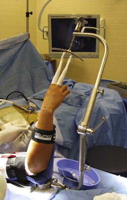
 A sterile traction tower is used to suspend the elbow flexed at 90 degrees.
 The index and long finger are placed in finger traps, and a 10-lb weight is applied through the traction tower (Fig. 65-1).
 A gravity inflow system is used instead of the typical infusion pump system to help decrease the chance of disseminating infected fluid into the surrounding soft tissue structures.
 An arthroscope, 30-degree angled lens, camera, and full radius joint shaver setup are needed for this procedure.
Exposures
 Extremity exsanguination is generally contraindicated because of the joint infection. In some situations a tourniquet may be used to allow better visualization of the wrist joint. If a tourniquet is used, the arm is elevated to let the blood drain before tourniquet inflation.
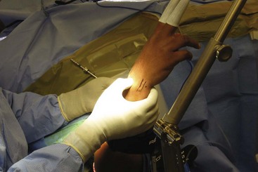
 The anatomic landmarks are identified before starting the procedure. The portal sites are then marked, which are the radiocarpal, 3-4, 6R, 6U, and radial and ulna midcarpal portals.
 The 6R portal is located by palpating the ECU tendon. The portal is made just radial to the tendon.
 The 3-4 portal is located by palpating the concavity between the extensor pollicis longus and EDC tendons just distal to Lister tubercle in line with the radial border of the long finger (Fig. 65-2).
 The radial midcarpal portal is located about 1 cm distal to the 3-4 portal, and the ulna midcarpal portal is located about 1 cm distal to the 6R portal in line with the midaxis of the ring finger.


