Procedure 62 Trapeziectomy and Abductor Pollicis Longus Suspensionplasty
![]() See Video 46: Abductor Pollicis Longus Suspension Arthroplasty for Basal Joint Arthritis
See Video 46: Abductor Pollicis Longus Suspension Arthroplasty for Basal Joint Arthritis
Examination/Imaging
Clinical Examination
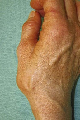
 The patient’s thumb metacarpal base may exhibit a dorsoradial prominence secondary to subluxation from ligamentous laxity (Fig. 62-1).
 The patient’s thumb should be evaluated for tenderness along the TMC joint.
 The carpometacarpal (CMC) grind test is performed with axial compression, flexion, extension, and circumduction of the TMC joint, which should elicit crepitus and pain.
 The patient’s thumb metacarpophalangeal (MCP) joint should be assessed during pinch for hyperextensibility. If unaddressed, the suspensionplasty will be stressed by obligatory metacarpal adduction. This hyperextensibility can be corrected with MCP joint capsulodesis or fusion if the deformity is severe.
 The patient should be evaluated for carpal tunnel syndrome, which may coexist in up to 43% of patients with TMC arthritis. Concomitant carpal tunnel release should be performed in these patients because trapeziectomy alone does not sufficiently increase carpal tunnel volume.
 An Allen test should be performed because the procedure typically requires mobilization of and potential injury to the radial artery.
 The patient should also be evaluated for concomitant trigger thumb.
Imaging
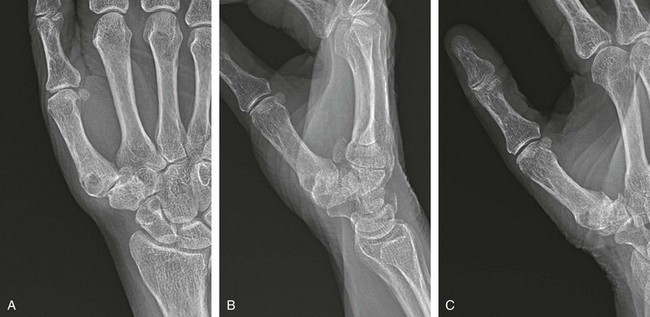
 Posteroanterior, oblique, and lateral radiographs of the hand should be obtained to evaluate the extent of arthritis, osteophyte formation, joint loss, and joint subluxation (Fig. 62-2A to C).
 The Eaton classification is a staging system based on radiographic findings only and often does not correlate with the patient’s symptoms. Some patients with severe arthritis may have minimal pain. Treatment should be based on the patient’s symptoms and, most important, on the amount of pain in the arthritic joint.
 A stress view of the TMC joint is obtained by having the patient press the thumb tips together while obtaining a 30-degree posteroanterior view centered on the thumbs to assess joint space loss and subluxation. Stress view is only obtained in stage I disease for patients with symptomatic joint laxity.
 Partial resection of the trapezoid should be performed if degenerative changes are present between the trapezoid and the scaphoid.
Surgical Anatomy
 The anterior oblique (beak) ligament inserts volarly onto the trapezium from its confluence with articular cartilage on the beak of the thumb metacarpal.
 The radial artery traverses the anatomic snuffbox and overlies the dorsal capsule of the CMC joint.
 The abductor pollicis longus (APL) and extensor pollicis brevis (EPB) tendons are within the first extensor compartment.
 The radial sensory nerve becomes subcutaneous after exiting through the interval between the brachioradialis and extensor carpi radialis longus (ECRL) muscles.
 The flexor carpi radialis (FCR) tendon traverses the volar and ulnar aspect of the trapezium.


