Fig. 1
(a, b) Pre-operative photograph demonstrating significant bilateral clubfoot and scars from posteromedial release

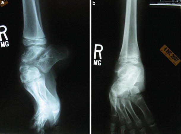
Fig. 2
(a, b) Pre-operative radiograph demonstrating midfoot and hindfoot deformity of the right foot. Lateral view of a right foot (a) and AP view of a right foot (b)
3 Preoperative Problem List
Midfoot
Varus
Supination
Equinus
Hindfoot
Equinus
Varus
4 Treatment Strategy
The pre-operative plan included left foot double osteotomy with a two-level application of the Biomet multiaxial correcting (MAC) external fixation system. The right was treated with no osteotomies and gradual correction with a two-level MAC system. The duration of the surgical procedure was approximately 2 h and the procedures were done 2 months apart.
5 Basic Principles
The use of the MAC external fixator allows for the correction and adjustment of angular deformity and displacement in all three planes, including the correction of residual or secondary deformities that occur during lengthening. Correction could be accomplished here with two MAC fixators, one on the hindfoot bisector medially and the other on the midfoot bisector, connected through a posterior 2/3 arc. The deformities can be corrected via a CORA-centric or CORA-perpendicular application. Correction can be accomplished through soft tissues if the joints are intact (Davidson 2011


Stay updated, free articles. Join our Telegram channel

Full access? Get Clinical Tree


