Figure 22.1
Tinea versicolor, hypopigmented round-oval flat lesions
Clinical Differential Diagnosis
(Pigmentation changes with minimal erythema)
Vitiligo
Post inflammatory hypopigmentation
Idiopathic guttate hypomelanosis
Tinea versicolor
Histopathology
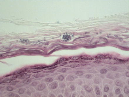
Sections showed minimal inflammation in the dermis. Within the cornified layer, numerous small yeast like organisms and hyphae structures were found by routine H&E (Fig. 22.2) or by KOH skin scraping prep (Fig. 22.3).


Figure 22.2


H&E 400×, Tinea versicolor showing numerous fungal yeast and hyphae forms found in the cornified layer
Stay updated, free articles. Join our Telegram channel

Full access? Get Clinical Tree


