Fig. 1
Pre-operative full-length standing radiographs revealing isolated distal tibia deformity and mild arthritis of the tibiotalar joint
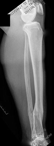
Fig. 2
Pre-operative lateral radiograph revealing posterior translation of the distal fragment and mild tibiotalar joint arthritis
3 Preoperative Problem List
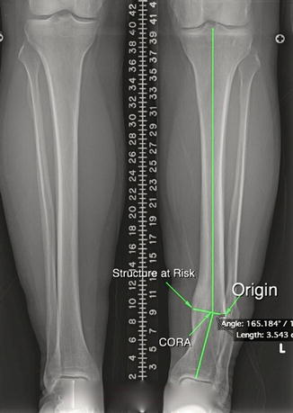
Deformity analysis (Fig. 3):
15° varus deformity
10 mm posterior translation of the distal fragment
7 mm shortening compared to contralateral side
5 degrees of external rotation (assessed clinically)
CORA at the level of her healed fracture
Metabolic issues:
Hypothyroidism
4 Treatment Strategy
Given the degree of deformity combined with shortening and rotation, the Taylor Spatial Frame was chosen to gradually correct the deformity. An acute correction could have been performed with internal fixation; however, this would have left the patient almost 1 cm short based on her deformity analysis. A gradual correction, restoring anatomic length, alignment, and rotation, would give her the best possible outcome.
5 Basic Principles
Related posts:
 8: Femoral Bone Defect
 11: Bone Transport Over a Nail for Infected Tibial Nonunion and Bone Defect
 22: Bone Transport to a Knee Fusion and Secondary Intramedullary Nailing s/p Gunshot Wound
 78: Two-Stage Treatment of a Chronic Foot Dislocation
 84: Lapidus Fusion with External Fixation
 65: Closed Correction of Club Foot with Ilizarov
Stay updated, free articles. Join our Telegram channel

Full access? Get Clinical Tree


