Procedure 52 Hemi-Hamate Arthroplasty
Figures 52-1, 52-5, 52-7, and 52-8 through 52-11 borrowed with permission from Williams RMM, Kiefhaber TR, Sommerkamp TG, et al. Proximal interphalangeal fracture/dislocations using a hemi-hamate autograft. J Hand Surg [Am]. 2003;28:856-865.
Indications
 Unstable PIP joint fracture dislocations in which more than 50% of the palmar base of the middle phalanx is fractured. There must be dorsal cortical continuity.
 Comminuted lateral plateau fractures of the base of the middle phalanx.
 Joint salvage after failed treatment of complex fracture-dislocations of the PIP joint.
Examination/Imaging
Clinical Examination
 Record active range of motion of the affected finger as tolerated.
 Evaluate coronal plane alignment, assessing for lateral deviation that suggests asymmetrical compression of the articular surface.
 Examine the sagittal alignment with the finger extended, looking for colinearity of the proximal phalanx and middle phalanx.
Imaging
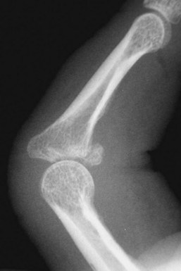
 Standard anteroposterior, oblique, and lateral views of the involved digit to assess the amount of articular involvement of the base of the middle phalanx, the integrity of the middle phalangeal dorsal cortex, and other fractures (Fig. 52-1).
 Computed tomography can better delineate the extent of articular cartilage involvement and comminution, but it is rarely necessary.
Surgical Anatomy
 The PIP joint is a complex hinge joint formed by the head of the proximal phalanx and the base of the middle phalanx that moves through a 110-degree arc of motion.
 The volar plate originates from the proximal phalanx periosteum and inserts onto the volar lip of the middle phalanx.
 Lateral joint stability is afforded by the thick collateral ligaments. The proper collateral ligament inserts onto the volar-lateral third of the middle phalanx, and the accessory collateral ligament inserts onto the volar plate.
 Dorsal translation of the middle phalanx is prevented by the volar plate and the cup-shaped articular surface of the middle phalanx, which congruously articulates with the proximal phalanx head.
 Dorsal fracture-dislocations disrupt both of these restraints and result in dorsal subluxation of the middle phalanx.
 The stability of the PIP joint is directly related to the amount of middle phalangeal volar articular surface disrupted. Fractures with as little as 30% of the articular surface involved may be unstable.
Exposures
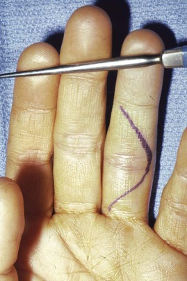
 Exposure of the PIP joint is gained through a palmar V-shaped incision centered over the PIP joint (Fig. 52-2).
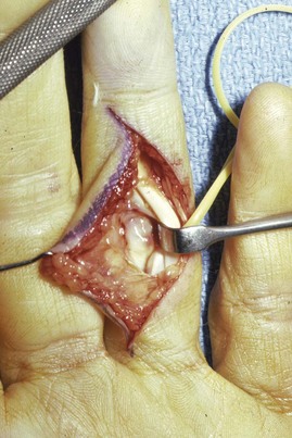
 The skin and subcutaneous tissue are sharply elevated, and both neurovascular bundles are visualized deep to the lateral digital sheet and protected.
 A rectangular flap of the fibro-osseous sheath that includes the entire A3 pulley is developed between the A2 and A4 pulleys.
 The flexor tendons are retracted to expose the volar plate, which is released from the accessory collateral ligaments at its lateral margins (Fig. 52-3).
 The volar plate is incised transversely from the base of the middle phalanx and reflected proximally (Fig. 52-4).
 Release the collateral ligaments from the head of the proximal phalanx, leaving a small stump attached to the middle phalanx to facilitate volar plate reattachment at the end of the procedure.
Stay updated, free articles. Join our Telegram channel

Full access? Get Clinical Tree


