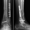
Fig. 1
AP view of both ankle joints before distraction. Lateral view is missing
3 Preoperative Problem List
Painful osteoarthritic ankle joint in a 45 year old man reporting a VAS score of 7–8 during standing and limiting the walking distance to 1 km.
Physical examination of the right ankle joint showed painful swelling and a range of motion of 0-5-45° which at the left side appeared to be 15-0-60°. No signs of ligamentous instability were found.
4 Treatment Strategy
In order to get the right foot into a plantigrade position, at first an arthroscopic debridement of the right ankle was done with removal of osteophytes on the distal tibia and talus without any surgery on the cartilage. A tibial frame consisting of two rings connected by screw-threaded rods was attached to a U-shaped foot ring having a half ring at the front. Two K-wires (1.8 mm in diameter), one from the posteromedial and one from the posterolateral side, were drilled through soft tissue and bone distally to the knee joint and another two proximally to the ankle joint. After securing of one end of a wire to the ring, these were fixed to the rings after being tensioned with 130 kN. Two K-wires with an olive were drilled from the medial and lateral side through the calcaneus under an angle of 30°. These wires were tensioned (0.9 kN) and fixed to the half ring around the heel. One pin with olive was drilled through the metatarsals I, IV, and V and tensioned to a half ring over the forefoot (0.5 kN). One K-wire was introduced through the talus and fixed to the half ring after being tensioned by 10 KN. Both the foot and distal tibia rings were connected by lengthening rods which allowed controlled distraction of talus from the tibia. To study the in vitro effects of low physiological levels of intermittent fluid pressure on unloaded articular cartilage possible (Van Valburg et al. 1998), a catheter with a pressure-sensitive tip was introduced into the ankle joint, tunneled subcutaneously, and connected to a measuring device. Distraction was carried out over a distance of 5 mm (0.5 mm twice daily for 5 days), starting the day after application of the apparatus. The patient was allowed to walk within a few days after surgery. After 12 weeks the external fixation apparatus was removed under general anesthesia. Full weight bearing was permitted.
5 Basic Principles
Levels of intermittent fluid pressure varied from 0 to 13 kPa (0.33 Hz), which were found to have beneficial effects on joint tissue in OA, indicating that this factor could be useful in treatment of OA (Van Valburg et al. 1998).
Related posts:
 8: Femoral Bone Defect
 11: Bone Transport Over a Nail for Infected Tibial Nonunion and Bone Defect
 30: C3.3 Pilon Fracture Closed. Ilizarov Fixation with Limited Open Reduction of Joint Surface and Distal Tibia Bridging Distraction of Ankle Joint
 78: Two-Stage Treatment of a Chronic Foot Dislocation
 84: Lapidus Fusion with External Fixation
 65: Closed Correction of Club Foot with Ilizarov
Stay updated, free articles. Join our Telegram channel

Full access? Get Clinical Tree


