Figure 29.1
Candidiasis, whitish material attached to the tongue (thrush)

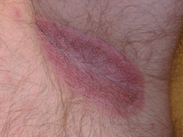
Figure 29.2
Candidiasis, beefy red plaque at the axillae
Clinical Differential Diagnosis
(Skin Fold Rash)
Intertrigo
Tinea
Psoriasis
Erythrasma
Seborrheic dermatitis
Candidiasis
Histopathology
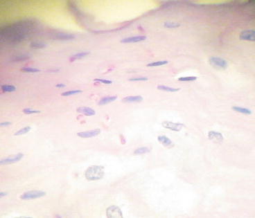
The changes of an infection can include an irregular psoriasiform hyperplasia with an overlying serum crust with neutrophils. The dermis shows a mixed infiltrate of histiocytes, lymphocytes, plasma cells, and occasional giant cell. The PAS stain shows yeasts (Fig. 29.3).


