Fig. 1
(a, b) X-rays showing post-traumatic arthrosis of the left ankle. Note ankle joint space narrowing especially in the anterior third of the joint

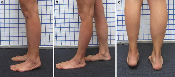
Fig. 2
(a–c) Side and back views showing equinus contracture. (a, c) Patient is unable to get heel to floor; (b) with left foot forward, heel touches the floor
3 Preoperative Problem List
1.
Post-traumatic ankle arthrosis
2.
Equinus contracture of ankle
4 Treatment Strategy
1.
Joint preservation approach. Avoid ankle fusion or ankle replacement in this young patient with ankle mobility.
2.
Anterior ankle arthrotomy, anterior debridement of scar tissue and osteophyte, and microfracture of areas of joint with full-thickness cartilage loss.
3.
Bone marrow aspiration from the ipsilateral iliac crest.
4.
Gastroc-soleus recession.
5.
Ankle distraction with circular hinged external fixator.
5 Basic Principles
1.
Ankle distraction leads to formation of reparative tissue in the ankle that increases the joint space on X-ray.
2.
Microfracture and addition of mesenchymal stem cells lead to formation of reparative tissue.
3.
Bone marrow aspirate concentrate (BMAC) contains mesenchymal stem cells.
4.
The ankle axis of rotation is oblique in both the coronal and axial planes. It is a line from posterolateral to anteromedial.
6 Images During Treatment
See Figs. 3, 4, and 5.

Get Clinical Tree app for offline access

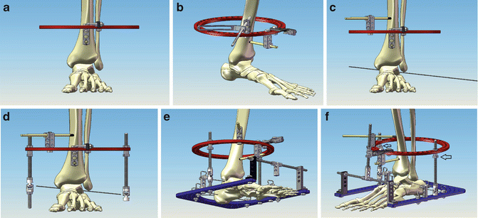
Fig. 3
(a–f) Schematic images of the SBi RingFIX RADTM frame showing surgical technique. (a) The tibial ring is applied orthogonal to the tibial axis. The location is 10 cm proximal to the ankle. (b) Stabilization of the tibial ring is with two 6 mm half-pins. (c) The axis of rotation of the ankle is located by inserting a trans-malleolar wire through the talus. (d


Stay updated, free articles. Join our Telegram channel

Full access? Get Clinical Tree


