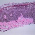
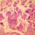
Figure 15.1
Mycobacterium tuberculosis
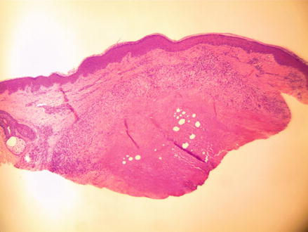
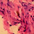
Figure 15.2
H&E 40× discrete subcutaneous nodule
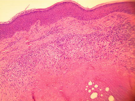
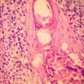
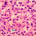
Figure 15.3
H&E 100×, outer wall consists of lymphocytes and histiocytes while the center is necrotic
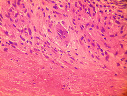
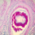
Figure 15.4
 6 Year Old White Male with Recurrent Brownish Patches Around Mouth
 37 Year Old White Male with Multiple, Brown, Purple Nodules on Arms and Legs
 70 Year Old White Male with Grouped Blisters on Right Lower Leg
 7 Year Old Male with an Enlarging, Round Area of Hair Loss
 29 Year Old White Female with Grouped Blisters on Left Thigh
 48 Year Old Hiv + Male with Multiple Papules and Nodules on Arms


H&E 400×, scattered multinucleate giants cells are found within outer wall
Related posts:
6 Year Old White Male with Recurrent Brownish Patches Around Mouth
37 Year Old White Male with Multiple, Brown, Purple Nodules on Arms and Legs
70 Year Old White Male with Grouped Blisters on Right Lower Leg
7 Year Old Male with an Enlarging, Round Area of Hair Loss
29 Year Old White Female with Grouped Blisters on Left Thigh
48 Year Old Hiv + Male with Multiple Papules and Nodules on Arms
Stay updated, free articles. Join our Telegram channel
Full access? Get Clinical Tree
