Figure 1.1
Bowenoid Papillosis arising in Condyloma acuminate
Clinical Differential Diagnosis
(Multiple red-brown, pigmented papules or plaque on genital skin)
Junctional Nevus
Seborrheic keratosis
Psoriasis
Bowenoid papulosis arising adjacent to condylomata acuminata
Condyloma lata
Histopathology
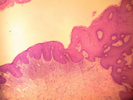
Microscopically, there were two distinct areas (Fig. 1.2). In one area, the epithelium was markedly hypertrophic with an overlying low papillomatous surface pattern while the adjacent epithelium was only slightly thickened compared to the surrounding epithelium. At higher magnification, there were additional distinctive features, the area that was less hypertrophic showed marked crowding of the keratinocytes with mitotic figures seen at all levels of the epithelium, while the thicker areas showed only a few mitotic figures with extensive koilocytic changes (keratinocytes with perinuclear clearing) at the upper layers of the epithelium (Figs. 1.3 and 1.4).


Figure 1.2


H&E 40×, Bowenoid papulosis on the left, while condyloma acuminata is on the right of this photo
Stay updated, free articles. Join our Telegram channel

Full access? Get Clinical Tree


