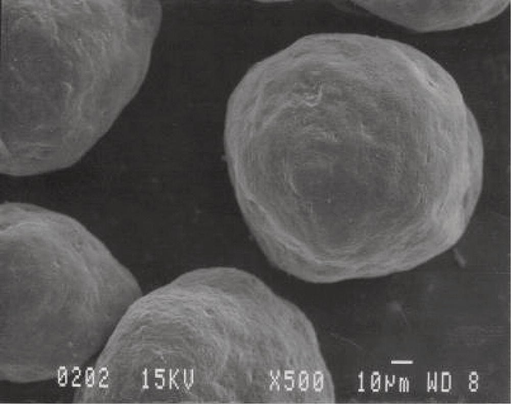
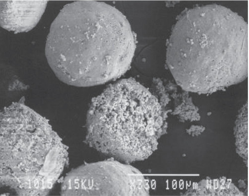
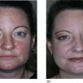
Stephen Mandy University of Miami Phillip Frost Department of Dermatology and Cutaneous Surgery, Miami, FL, USA Facial volume loss leads to dramatic changes in appearance, and may result from aging, disease, or hereditary conditions. The deflation from lipoatrophy causes skin redundancy, which is compounded by loss of elasticity and collagen degeneration as a consequence of solar radiation and oxidative damage. Skeletal resorption further leads to deflation, enlargement of the ocular orbit, and shrinkage of the jaw. These visual signs of aging cannot be corrected by surgical tightening without volumization as it will lead to a skeletal, windswept appearance. Replacement of volume through soft tissue augmentation can often offer facial rejuvenation with or without surgery. A variety of suitable materials for soft tissue augmentation exists. Natural fillers, such as collagen, hyaluronic acid (HA), and calcium hydroxylapatite (CaHA), are synthesized to mimic, or are derived from, naturally occurring biological materials. Synthetic fillers may be permanent, such as acrylates, silicone, or biodegradable such as poly‐L‐lactic acid [1]. CaHA is a type of fibroplastic filler in which volume correction is achieved in part through the biological response of the host. Radiesse® (Merz Aesthetics, Raleigh, NC) is an injectable filler composed of microspheres of CaHA (30%) suspended in an aqueous gel consisting of water, glycerin and sodium carboxymethylcellulose. Once injected, the gel carrier is soon absorbed. The microspheres are 25–45 μm in diameter, which facilitates injection, but resists immediate phagocytosis. These bioceramic spheres have no antigenicity, foreign body or giant cell response, and cause a minimal inflammatory reaction. They do not stimulate ossification. Though visible on X‐ray images and magnetic resonance imaging scans, they are radiolucent, appearing somewhat like frosted glass, and pose no impediment to radiological analysis. The CaHA microspheres form a scaffold for the fibroplastic proliferation, which provides the natural tissue feel of the implant. The local fibroplastic response results in the fibrous encapsulation of the particles and their gradual dissolution into calcium and phosphate ions. (Figures 46.1–46.3) [2, 3]. In a recent study, biopsies performed 6 months following injection of CaHa revealed an increase in elastic fibers between 30% and 80% and an increase in Proteoglycans between 12% and 77% [4]. A similar study comparing CaHa and HA revealed that CaHa produced a highly significant increase in Type 3 collagen at 4 months which was then replaced by Type 1 collage at 9 months [5]. These studies validate the improvement in skin quality seen with the injection of CaHa. Figure 46.1 Calcium hydroxylapatite microspheres, 25–45 μm in diameter. (Source: Illustration courtesy of BioForm Medical.) Figure 46.2 Gradual dissolution of microspheres. (Source: Illustration courtesy of BioForm Medical.) CaHA is a thick, white, clay‐like, cohesive material intended for subdermal injection. Radiesse L®
CHAPTER 46
Calcium Hydroxylapatite for Soft Tissue Augmentation
Introduction
Physiology and Pharmacology
Indications and techniques
Related posts:
![]()
Stay updated, free articles. Join our Telegram channel


Full access? Get Clinical Tree


