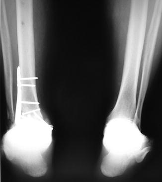
Fig. 1
(a, b) Pre-operative AP X-ray with distal tibia intra-articular nonunion, ankle post-traumatic arthritis, and retained hardware

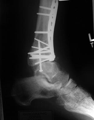
Fig. 2
Pre-operative lateral X-ray with anterior distal tibia bone erosion and talar subluxation

Fig. 3
Bilateral hindfoot alignment views
3 Preoperative Problem List
1.
Distal tibia intra-articular nonunion
2.
Post-traumatic ankle arthritis
3.
Bone loss of the distal tibia
4.
Ankle joint subluxation with anteriorly displaced talus
5.
Diabetes type II
6.
Osteopenia
7.
Obesity
4 Treatment Strategy
To restore a stable plantigrade limb, the treatment plan consisted of distal tibia and fibula resection, deformity correction, and ankle arthrodesis. Approximately 2.5 cm of bone at the distal tibia was resected. Limb shortening was achieved acutely without neurovascular compromise. Ankle arthrodesis was performed acutely with internal fixation. Ring external fixation methods were used to supplement the fusion stability and restore limb length. A proximal tibia osteotomy was performed using the Gigli saw technique, and a fibular shaft osteotomy was performed with a sagittal saw. After a 7-day latency period, lengthening (0.75 mm/day) using Taylor Spatial Frame methods was completed. The frame was removed after regenerate consolidation and arthrodesis healing.
5 Basic Principles
Limb deformity, joint integrity, nonunion physiology, and host state (Cierny et al. 1985) were evaluated pre-operatively with radiographs, CT scan, clinical history, and laboratory results (CBC, ESR, CRP, metabolic panel, vitamin D, HgbA1C, and thyroid function).
Bone resection and primary arthrodesis were chosen due to joint destruction, bone loss, and intra-articular nonunions with nonviable bone segments. Limb salvage and limb-length restoration was chosen in this high-functioning and relatively young patient despite several stable medical issues.
Stay updated, free articles. Join our Telegram channel

Full access? Get Clinical Tree


