Figure 3.1
Lichen Planus
Clinical Differential Diagnosis
Drug eruption
Lichen Planus secondary to Hepatitis C infection
Psoriasis
Tinea
Contact dermatitis
Histopathology
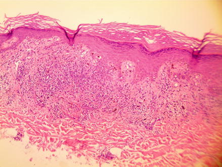
Sections showed a band-like infiltrate of lymphocytes along the epidermal/dermal junction. The junction showed “sawtooth” like changes. Focal wedge shaped hypergranulosis was seen (Fig. 3.2) Period Acid-Schiff stain tested negative for fungus.


Figure 3.2


H&E 100×, Lichen planus eruption secondary to Hepatitis C infection
Stay updated, free articles. Join our Telegram channel

Full access? Get Clinical Tree


