Fig. 1
Pre-operative radiographs: malunion of the left femur in its upper third – translation of the bone fragments in two planes, shortening and varus angulation observed. MAD = 11 mm medial, mLPFA = 90°, mLDFA = 87°, mMPTA = 87°, mLDTA = 91°
3 Preoperative Problem List
(a)
Malunion of the left femur accompanied by complex four-component, two-plane deformity
(b)
Shortening of the left femur, 3 cm
4 Treatment Strategy
Our plan is chosen according to the complexity of the deformity, the character of the initial damage (gunshot injury), the risks of infectious complications, and the time since the fracture occurred. The plan is for external fixation of the femur, using gradual deformity correction with the computer-assisted Ortho-SUV Frame.
5 Basic Principles
(a)
Use of external fixation provides possibility of gradual lengthening and deformity correction
(b)
Possibility of closed procedure decreases the invasiveness of the treatment and the risks of infectious complications
(c)
Use of computer-assisted hexapod provides precise gradual deformity correction
6 Images During Treatment
See Figs. 2, 3, 4, 5, 6.


Get Clinical Tree app for offline access

Fig. 2
Photos and radiographs after the surgery: Ortho-SUV frame is applied

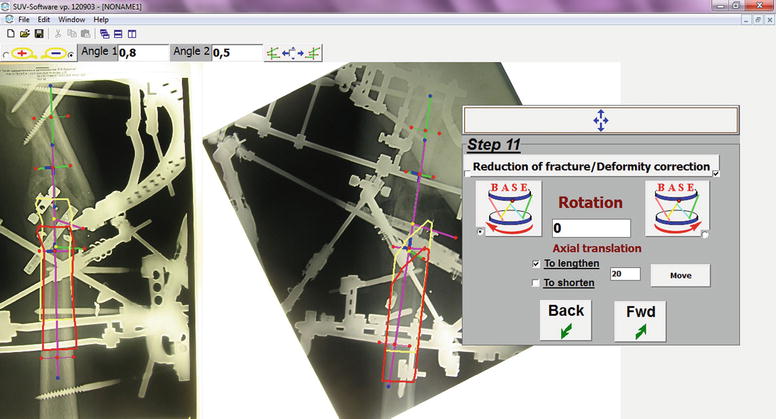
Fig. 3
The Ortho-SUV software window at step 11, the deformity correction planning. First step involves lengthening over the axis of the distal bone fragment to avoid bone fragment collision during deformity correction. Yellow bone contour indicates the initial mobile (corresponding) fragment position. Red bone contour demonstrates the position of the mobile fragment after deformity correction is achieved

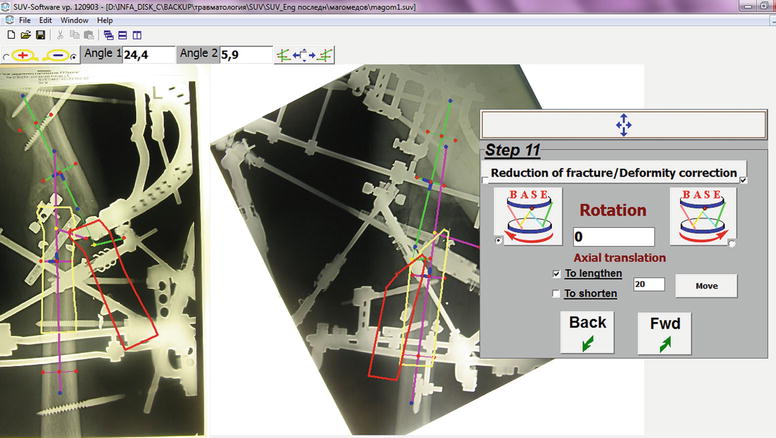
Fig. 4
The Ortho-SUV software window at step 11, the deformity correction planning. Second step –final planning of deformity correction (total residual). The bone fragments anatomical axes are drawn. Yellow


Stay updated, free articles. Join our Telegram channel

Full access? Get Clinical Tree


