Fig. 1
(a, b) Pre-operative clinical pictures with shortening of the right lower limb and forefoot deformity
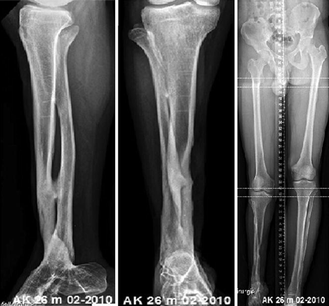
Fig. 2
Pre-operative X-rays
3 Preoperative Problem List
1.
Post-traumatic shortening of the lower limb
2.
Pes equinus deformity of the fused ankle joint and varus deformity of the forefoot
4 Treatment Strategy
1.
Lengthening of the lower leg
2.
Axis correction by osteodistraction, using a multiplanar ring fixator
3.
Acute correction of the malaligned fused ankle joint
5 Basic Principles
Distraction osteogenesis has been widely used for the treatment of leg-length discrepancy, nonunion, traumatic bone defects, complex deformities, and osteomyelitis. In this case, the proximal tibia was osteotomized, and limb lengthening was performed with a circular ring fixator.
6 Images During Treatment
See Figs. 3, 4, 5, 6, and 7.
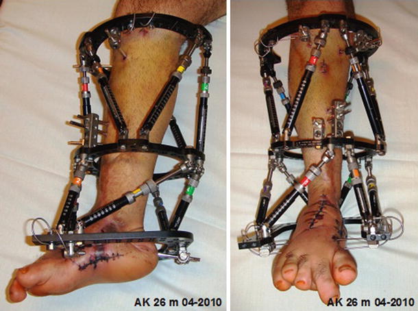
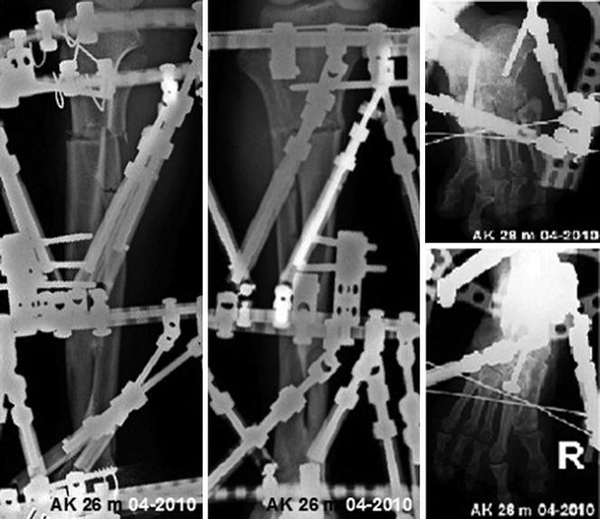
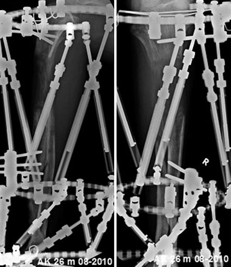
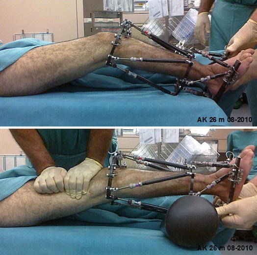
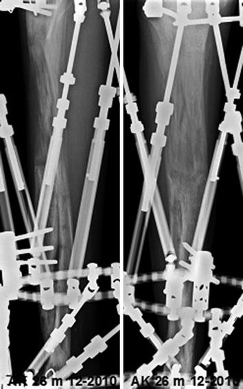
Fig. 3
Hexapod ring fixator for tibial lengthening and stabilizing the acute forefoot correction
Fig. 4
Post-operative X-rays
Fig. 5
X-rays 18 weeks post-op
Fig. 6
Surgical arthrolysis of a knee joint contracture was necessary in this case
Fig. 7
Thirteen months post-operatively
7 Technical Pearls
Related posts:
 8: Femoral Bone Defect
 11: Bone Transport Over a Nail for Infected Tibial Nonunion and Bone Defect
 22: Bone Transport to a Knee Fusion and Secondary Intramedullary Nailing s/p Gunshot Wound
 78: Two-Stage Treatment of a Chronic Foot Dislocation
 84: Lapidus Fusion with External Fixation
 65: Closed Correction of Club Foot with Ilizarov
Stay updated, free articles. Join our Telegram channel

Full access? Get Clinical Tree


