Figure 10.1
Kaposi sarcoma presents as a brownish purple nodule
Clinical Differential Diagnosis
Lymphoma
Leukemia
Metastatic malignancy
Angiosarcoma
Kaposi sarcoma
Infection
Histopathology
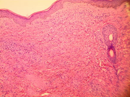
A 4 mm punch biopsy was performed on the superior left arm. Sections show an ill-defined lesion comprised of short spindled cells. Some of these cells form irregular vascular channels that encircle existing vessels (promotory sign) (Fig. 10.2). While some vessels contain RBCs, other areas show extravasated RBCs with hemosiderin deposits (Fig. 10.3).


Figure 10.2


H&E 100×; Kaposi sarcoma is caused by the HHV8 virus. Multiple slit like vascular channels are found
Stay updated, free articles. Join our Telegram channel

Full access? Get Clinical Tree


