Fig. 1
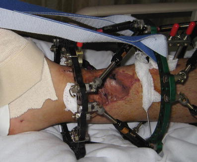
There is a large, medial, open wound with an antibiotic cement spacer protruding

Fig. 2
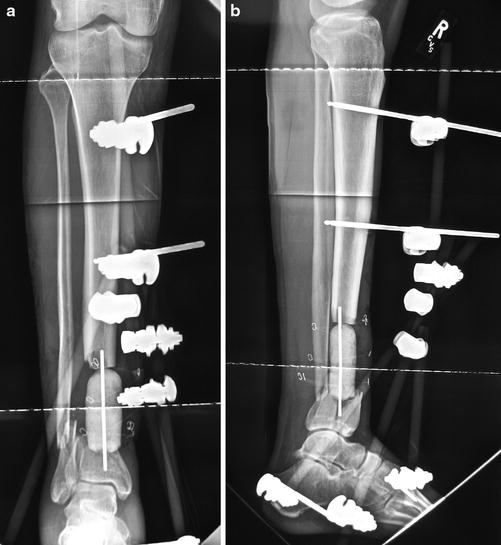
(a, b) AP and lateral radiographs show the provisional pin-to-bar frame and cement spacer. There is a coronal split of the short, distal fragment with a nondisplaced articular component
3 Preoperative Problem List
1.
Distal tibial bone defect
2.
Distal medial leg soft tissue defect
3.
Coronal intraarticular fracture
4 Treatment Strategy
The plastic surgery team was consulted, and a discussion of all options ensued. Options included bone transport and local grafting, free tissue and free fibula transport, and below-knee amputation. The patient was included in the decision and bone transport was selected.
5 Basic Principles
The wound was debrided, spacer removed, articular surface fixed, external fixator applied, and a local soleus flap mobilized and covered with a split-thickness skin graft. The grafts were allowed 4 weeks to heal, and then a two-level tibial osteotomy was performed. The bone transport was started with the proximal site distracted at 0.75 mm per day, the mid-tibial osteotomy site distracted 0.5 mm per day, and the defect site shortened at 1.25 mm per day.
6 Images During Treatment
See Figs. 3, 4, 5, 6, and 7.


Fig. 3
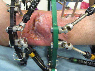
The wound has been covered and the transport has begun

Fig. 4
The transport segment is entrapping the overlying skin graft into the docking site. This required surgery for skin elevation and docking site grafting. The use of this technique avoided the need for free soft tissue transfer with microvascular anastomosis
Stay updated, free articles. Join our Telegram channel

Full access? Get Clinical Tree


