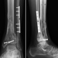
Fig. 1
(a) Anteroposterior radiograph at time of referral to our institution. (b) Lateral radiograph corresponding to Fig. 1a
3 Preoperative Problem List
1.
Nonunion/malunion of distal tibia and fibula
2.
Painful fibular hardware
3.
History of tibia infection
4 Treatment Strategy
At the time the patient presented to us, his tibial incision where a plate was placed and subsequently removed was well healed. It showed no signs of active infection . The plan was to remove the fibular plate, apply a spatial frame to match the deformity, and start a gradual correction . An osteotomy of the fibula was performed in order to permit easier tibial correction. The fibular osteotomy was made proximal to the ankle syndesmosis which corresponded to the center of the tibial deformity (Fig. 3a). Given the history of wound issues, the goal was to correct the deformity and avoid further hardware placement if possible after frame removal. The primary goal was to correct the varus deformity. Consideration was given to more correction of the sagittal plane deformity, but this would have required soft tissue releases posteriorly.
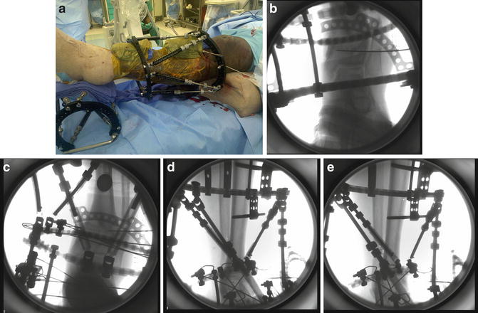
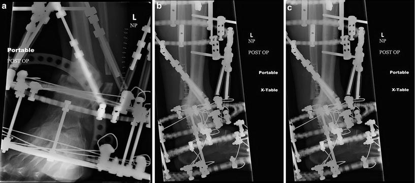
Fig. 2
(a) Intra-operative photo demonstrating one method of centering the leg for frame application purposes. Note the folded towels held to the leg anteriorly with a sticky drape. (This image is from a different patient but included to illustrate frame application tips). (b) Intra-operative fluoroscopy image demonstrating reference wire applied parallel to ankle joint and perpendicular to tibial anatomic axis. (c) Intra-operative fluoroscopy image demonstrating reference sphere in the image for measuring mounting parameters post-operatively. The skinny wire proximal to the ankle is not a mounting reference wire but now serves to identify the deformity origin or center of rotation of angulation (CORA). (d and e) Intra-operative fluoroscopy image. Note the improved overlap between the two posts on the ring proximal to the deformity. The ring is superimposed on itself in Fig. 2e as are the posts. This image now needs the measuring reference sphere, and then accurate mounting measurements can be completed
Fig. 3
(a) Post-operative radiograph of the ankle after frame application. Note difficulty in viewing the distal tibia. The fibular osteotomy is visible, and the gradual loss of height here indicates correction of the varus deformity. (b and c) Post-operative radiographs demonstrating two rings per bony segment and two or more points of fixation per ring
5 Basic Principles
Related posts:
 8: Femoral Bone Defect
 28: Proximal Tibial Bone Defect Treated with Intentional Deformity and Bone Transport
 30: C3.3 Pilon Fracture Closed. Ilizarov Fixation with Limited Open Reduction of Joint Surface and Distal Tibia Bridging Distraction of Ankle Joint
 78: Two-Stage Treatment of a Chronic Foot Dislocation
 84: Lapidus Fusion with External Fixation
 65: Closed Correction of Club Foot with Ilizarov
Stay updated, free articles. Join our Telegram channel

Full access? Get Clinical Tree


