Procedure 34 Reconstruction of the Central Slip with the Transverse Retinacular Ligament for Boutonnière Deformity
![]() See Video 26: Correction of Boutonnière Deformity
See Video 26: Correction of Boutonnière Deformity
Indications
 Boutonnière deformity is classified into three stages by Nalebuff, as follows:
 Indication: Boutonnière deformity of the PIP joint with almost full passive extension (stages 1 and 2) is suitable for this reconstructive surgery.
 Contraindication: If articular destruction is evident or if a severe fixed flexion contracture is present (stage 3), arthrodesis or arthroplasty of the PIP joint is recommended.
Examination/Imaging
Clinical Examination
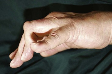
 Boutonnière deformity is seen in a patient with rheumatoid arthritis (Fig. 34-1).
 Passive PIP joint extension should always be evaluated before surgical reconstruction.
 The mobility of the PIP joint is very important for obtaining good results. Therefore, in the case of the boutonnière deformity with severe flexion contracture of the PIP joint, rehabilitation or joint splinting in extension of the PIP joint should be performed before the operation.
Imaging
 Radiographic examination is required to evaluate the status of the metacarpophalangeal (MCP), proximal interphalangeal (PIP), and distal interphalangeal (DIP) joints.
 If the PIP joint is destroyed, arthrodesis or arthroplasty is recommended.
 Magnetic resonance imaging examination may help to identify synovitis of the PIP joint.
Surgical Anatomy
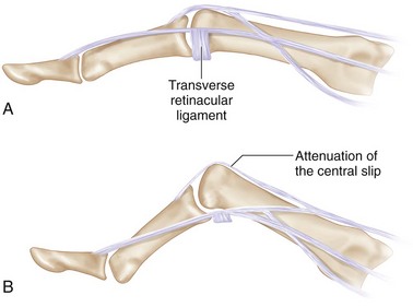
 Disruption of the central slip of the extensor tendon results in palmar migration of the lateral bands with PIP joint flexion. Proximal displacement of the extensor apparatus results in the DIP joint hyperextension deformity (Fig. 34-2A is normal; B shows boutonnière deformity).
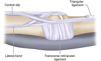
 The transverse retinacular ligament is the key component in this procedure. This ligament holds the lateral bands in the palmar direction during flexion. It inserts into the palmar plate of the PIP joint and flexor tendon sheath (Fig. 34-3).


